Breathing and supporting activity
The hummingbird has left the flower and settled on a branch. Its wings are folded, but air continues to enter and leave its body. If we could follow that air, we would not find a miniature version of a human lung. We would find a system in which flexible air sacs help ventilate comparatively rigid lungs, and gas exchange occurs in finely divided lung tissue. The conspicuous spaces and the principal exchange surfaces have different jobs.
This distinction is the center of the chapter. Breathing moves air. Gas exchange moves molecules across a barrier. Circulation moves blood. Cellular metabolism uses oxygen in processes that help make chemical energy available. These events are connected, but one cannot substitute for the others. A correct explanation must carry oxygen from the environment to a working cell without skipping a necessary step.
Separate the ventilator from the exchanger
The large air sacs are thin-walled spaces connected to the respiratory passages. They change volume as the body moves during breathing. The lungs contain branching airways, including parabronchi, with minute air spaces closely associated with blood capillaries. The air sacs primarily serve ventilation; they are not the principal sites of oxygen transfer into blood. In the main paleopulmonic region, air passes through parabronchi in the same general direction during both respiratory phases. Maina, 2022: structure and function of the avian respiratory system.
A pair of bellows and a separate exchange unit offers a limited analogy. Changing the bellows' volume can drive flow through another structure. But a bird does not contain leather bellows, and a diagram should not invent mechanical flap valves simply because they make a familiar machine work. Airway geometry and flow dynamics matter in the actual animal.
This separation helps explain a misleading impression from anatomical drawings. The air sacs often occupy much more of the picture than the lung tissue. Large visible volume does not establish large exchange capacity. Exchange depends on properties such as the relevant surface area, barrier thickness, gas gradients, and blood supply. A spacious chamber and a finely divided exchange region can have very different consequences despite both containing air.
The lungs are comparatively stable in shape while the volume of the surrounding respiratory apparatus changes. Birds do not ventilate by expanding mammalian alveoli with a mammalian diaphragm. This is a difference in arrangement, not permission to declare one animal's entire physiology superior to another's. Different bodies support demanding activities through different combinations of structures.
Follow one marked parcel without stopping the rest
The original schematic below tracks an imagined parcel of air through a simplified route. It is a teaching model, not a claim that the respiratory system handles sealed packets with no mixing. The words “first” and “second” identify successive phases for that marked parcel; other air is moving at the same time.
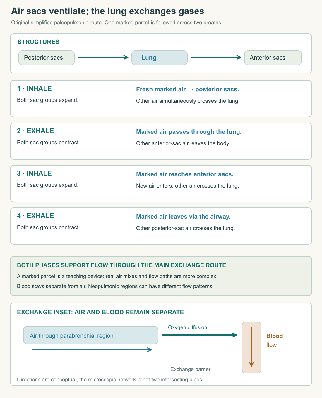
During the first inhalation in this simplified account, the marked air enters toward the posterior, or caudal, air sacs. During the following exhalation, those sacs supply air toward the lung's main through-flow pathway. The next inhalation draws the marked air onward toward the anterior, or cranial, sacs. The next exhalation carries it out. The model therefore takes two breathing cycles to show the marked parcel's full route.
The crucial correction is that the lung does not wait, empty and idle, while one parcel completes this journey. During inhalation, air can pass through the main exchange pathway toward the expanding anterior sacs while newly entering air supplies the posterior region. During exhalation, posterior sacs supply that pathway while anterior sacs discharge outward. A four-panel parcel sequence must be read alongside these concurrent movements.
Why bother with this qualification? Because a sequence drawn as four isolated boxes can accidentally teach that gas exchange occurs only on alternate breaths. The error comes from confusing the path of a marker with the state of the whole system. A commuter passing through four stations does not mean that all other commuters stop until that person's journey ends.
The station analogy has its own limit. Air mixes, flow is distributed through branching passages, and anatomical details vary. The schematic omits much of that detail so you can retain the main distinction between ventilation and exchange. In some bird lungs, a region called the neopulmo has different flow characteristics. The familiar unidirectional description should not be stretched into “all air always moves one way through every passage.”
A useful verbal check is to narrate the whole system during an inhalation without mentioning the marked parcel. Both groups of sacs expand, but their connections mean that the air entering them has followed different routes. Then narrate an exhalation: both groups decrease in volume, supplying different outflow routes. If your story requires one group to expand while the other contracts, you have changed the model into a different machine.
A directional pathway needs physical evidence
It would be easy to assume that one-way airflow requires a row of anatomical doors. That is an engineering guess. Experiments must establish whether the relevant direction can arise from the structure and flow itself.
Scheid, Slama, and Piiper studied fixed duck lung preparations separated from the air sacs and other organs. When pressure or suction ventilated the main bronchus, their reported dorsobronchial flow retained its direction toward the parabronchi in both phases. The abstract supports the importance of airway structure in directing flow; it does not mean an isolated preparation reproduces every condition in an actively breathing bird. Scheid and colleagues, 1972: study abstract.
Notice the logic. If a pattern remains in a preparation without the original living control system, some explanations become less necessary for that particular result. It does not follow that living control has no role anywhere in respiration. The experiment narrows a causal question; it does not abolish all the surrounding physiology.
Our schematic uses arrows instead of imagined flaps for that reason. An arrow is a claim about a route or direction. It should not quietly become a drawing of a structure that has not been established. This is the same discipline used in the flight chapter: a helpful visual must make its physical commitments explicit.
Oxygen crosses a barrier
Airflow brings gases near an exchange surface. Oxygen then moves by diffusion according to its partial-pressure gradient. Partial pressure describes the contribution of one gas to a mixture's pressure and provides the relevant language for gas exchange. Surface area, diffusion distance, and the gradient influence transfer. Oxygen and carbon dioxide each follow their own gradients; they are not exchanged as one-for-one tokens. OpenStax: principles of respiratory gas exchange.
Imagine two sealed regions separated by a barrier permeable to a particular gas. If a gradient initially favors movement from one side to the other, transfer tends to reduce that difference. To sustain substantial transfer, a system must help maintain suitable conditions on both sides. Ventilation refreshes respiratory gas, while blood flow carries gases to and from the exchange region.
This explains why “thin surface” is not a complete answer to how a lung works. A thin surface with no useful gradient cannot produce the same transfer as a thin surface with an appropriate gradient. A well-ventilated region with too little blood flow also cannot deliver unlimited oxygen to the body. The operation depends on matching several processes.
At the parabronchial scale, avian exchange is commonly described as crosscurrent: blood approaches the exchange region across the general direction of airflow. The microscopic network is more complicated than two straight pipes. Our drawing therefore shows a conceptual relationship, with a barrier between air and blood, rather than copying the countercurrent gill diagram from the fish course. Crosscurrent does not mean that air and blood pour into one shared tube.
Try reading the arrows as molecules instead of fluids. An oxygen molecule crosses from an air space through the intervening barrier into blood under appropriate conditions. The air itself does not bubble through the bloodstream. Later, oxygen leaves blood near tissues where conditions favor unloading and diffusion. Keeping these movements distinct prevents a surprisingly common confusion between circulation and ventilation.
Blood completes the delivery route
A bird's four-chambered heart supports pulmonary and systemic circuits. Blood returning from the body reaches the right atrium and right ventricle, then travels to the lungs. Blood returning from the lungs reaches the left atrium and left ventricle, then travels to the body's tissues. Arteries carry blood away from the heart and veins return it; those names do not mean “always oxygen-rich” and “always oxygen-poor.” OpenStax: circulatory organization.
This route adds a necessary step to the common sentence “the lungs send oxygen to the muscles.” The lung is an exchange site. Blood transports much of the oxygen, and the heart provides the pressure differences that sustain circulation. A concise sentence can remain useful, but a diagram needs to show the intervening route.
Hemoglobin in red blood cells binds oxygen reversibly. Binding greatly increases how much oxygen blood can carry compared with the amount dissolved alone. Reversible binding is essential: a carrier that never releases its load would not solve tissue supply. Carbon dioxide travels in several forms, including bicarbonate, rather than merely as bubbles returning to the lung. These are shared vertebrate transport principles; human numerical values should not be assigned to a hummingbird. OpenStax: gas transport mechanisms.
The distinction between amount and pressure matters here. Two blood samples need not carry identical total oxygen amounts merely because their oxygen partial pressures match. Hemoglobin concentration and binding behavior affect content. For this course, you do not need to calculate a full dissociation curve. You do need to avoid treating every oxygen measurement as the same quantity.
An original delivery model makes the relationship concrete. Suppose one unit of blood delivers two arbitrary oxygen units to a tissue, and three blood units pass per minute. The modeled delivery is six oxygen units per minute. Doubling flow could double delivery if the amount supplied per blood unit remained the same. If that amount fell at the same time, the result would differ. The invented numbers expose the assumptions behind the multiplication.
A faster heart rate is likewise not a complete measurement of flow. The amount moved per beat matters too. Nor does faster breathing alone establish how much oxygen reaches a muscle. Every visible rate sits within a chain of volumes, gradients, transport capacities, and tissue demand.
Supporting activity means coordinating a chain
Return to the hummingbird's departure from the branch. The wing muscles change their activity, but sustained performance also requires an adequate supply of usable fuel and oxygen. The respiratory and circulatory systems participate in that supply. They also carry consequences away, including carbon dioxide and heat.
It is useful to call this an oxygen transport cascade: environmental availability, ventilation, exchange, blood transport, tissue diffusion, and cellular use are linked stages. The word cascade does not mean one stage always limits performance. Which stage matters most can change with the species, the environment, and the intensity or duration of activity.
Imagine three hypothetical constraints. First, lower atmospheric pressure reduces the oxygen partial pressure of otherwise similar dry air. Second, a transport system carries less oxygen per unit blood. Third, a muscle requires oxygen at a higher rate. Each could alter the relationship between supply and demand, but they enter the chain at different places. A single response such as faster breathing may not compensate equally for all three.
The altitude example is worth stating carefully. The oxygen fraction in ordinary dry air remains approximately the same over common terrestrial altitude comparisons; the lower total pressure reduces its partial pressure. Saying “there is no oxygen up there” misses the relevant quantity. We will not assign a particular altitude tolerance to either focal species without species-specific evidence.
A bird also pays for ventilation. Moving the body wall and air through passages requires work. Large breaths and frequent breaths can impose different mechanical demands, and some air movement occurs in conducting spaces rather than at exchange surfaces. More movement is therefore not automatically a more economical way to supply tissues. The appropriate account asks what useful exchange results from the work.
What an outside observer can and cannot establish
A resting bird's body movements may permit a breathing count when the view is good. A distant, moving, or partly concealed bird may not. Feather motion, posture changes, and recording frame rate can complicate interpretation. If you cannot confidently distinguish breaths, do not convert ambiguous movement into a physiological statistic.
Even an accurate breathing count is only one variable. It does not provide the volume of each breath, gas composition, pulmonary blood flow, or tissue oxygen use. A short notebook entry can acknowledge that limit and still ask a useful question: does a visible respiratory movement change after spontaneous activity, under comparable viewing conditions?
For this course, observation never requires restraint, handling, or an induced flight. Published research and original paper models supply the evidence for internal mechanisms. Wildlife can continue its day while you improve your account of what a visible action does and does not reveal.
You should now be able to trace two routes without merging them. Air follows respiratory passages and changing air-sac volumes; blood follows vessels and the heart. Oxygen crosses between them at the lung's exchange tissue, then crosses toward working tissues elsewhere. Their coordination supports activity, but the anatomy alone does not establish the maximum performance of every bird.
The next chapter reconnects that delivery chain with food. Oxygen is part of aerobic metabolism; it is not a meal. The hummingbird and gull must obtain, process, and allocate suitable materials while keeping the resulting heat balance workable. Breathing makes more sense when placed back inside that larger working day.
A final diagram check is to follow air and blood with separate pencils. They approach an exchange barrier without becoming one circulating fluid. If an arrow pours air directly into a blood vessel, the schematic has removed the very surface whose diffusion function it is supposed to explain.
Application
Reconstruct the respiratory sequence from memory, then compare it with the original diagram. Use one color for an imagined marked air parcel and another for air already moving elsewhere. Add a separate loop for blood, label the four heart chambers in order, and mark the sites where oxygen crosses a barrier. Include a note explaining why the air sacs are not the main exchange surfaces.
Next, write a 300-word explanation of how a bird can maintain flow through the main parabronchial pathway during both inhalation and exhalation. Name two simplifications in your drawing and explain why the two-breath parcel model does not imply that the lung exchanges gas only every other breath.
Check your understanding: Does doubling the visible breathing frequency prove that twice as much oxygen reaches the wing muscles?
Expected answer: No. Breath volume, effective ventilation, gas gradients, exchange, blood flow, oxygen content and tissue uptake also matter. Frequency is one part of a coordinated transport chain, not a direct measurement of oxygen delivery to a particular tissue.