Signals and connections
A hand moves toward a cup, stops, and reaches for a different one. Nothing about the basic electrical event in a neuron has to become “cup-shaped” to support that change. The relevant information lies in activity across particular cells and connections, unfolding in time. To understand that statement, we need to follow a signal along an axon, through a synapse, and into a circuit. These are three related events, with three different explanatory jobs.
A changing membrane
An action potential is a brief, actively regenerated change in membrane voltage. It is not a packet of neurotransmitter moving down the axon. Nor is it a group of sodium ions traveling all the way from the cell body to the terminal. Ions cross the membrane locally; the electrical change affects neighboring membrane, where another local change can be generated. Propagation depends on the properties of the route it travels through.
Start with a simplified neuron whose inside is negative relative to outside. Depolarization makes that difference less negative. If depolarization activates enough voltage-sensitive sodium channels, inward sodium current can produce further depolarization and activate more channels. This regenerative process produces the rising phase of a typical neuronal spike. Its threshold is a condition for this self-amplifying event, not a universal voltage printed on every neuron.
The rise ends because sodium channels inactivate and other currents, especially outward potassium current in the standard introductory example, become more influential. Repolarization brings voltage back toward its resting level. A temporary afterhyperpolarization can make the inside more negative than its preceding resting value. The detailed waveform varies across cell types and conditions. A textbook curve represents a useful pattern, not an invariant electrical signature of all human neurons.
During an absolute refractory period, the membrane cannot produce another ordinary spike because crucial sodium channels have not recovered from inactivation. During a subsequent relative refractory period, producing another spike is more difficult than before. These changing conditions constrain timing. They also help explain why a normally initiated axonal signal proceeds toward the terminal rather than repeatedly exciting the membrane it has just passed.
The phrase all-or-none describes the regenerative character of the event in a given excitable membrane under specified conditions. It does not mean that the whole neuron has only two possible states, that all its signals are identical, or that every input either does everything or does nothing. Small changes in voltage can occur without a spike. Cells can differ in their firing patterns. Even spike shape can vary with circumstances. The useful claim is more specific: crossing threshold recruits a regenerative event rather than merely scaling up a small passive voltage change.
How a signal travels
Place two imaginary recording sites along one axon. In our fictional example, they are three centimeters apart, and the recognizable event appears at the second site two milliseconds after the first. Divide 0.03 meters by 0.002 seconds: the inferred speed is fifteen meters per second. This calculation concerns the movement of the electrical event between recording positions. It is not the speed at which a particular sodium ion traverses the axon's entire length.
The distinction resembles a line of people standing in sequence. A visible wave can move rapidly along the line even though each person moves only near their own position. This analogy captures propagation by local events. It does not explain the membrane channels, gradients, or refractory period; those need the biological account already given. An analogy should identify one useful relationship rather than claim to replace the mechanism.
Myelin changes propagation by limiting current leakage and altering how much charge must be moved to change voltage across the wrapped membrane. Local current spreads between nodes, where the action potential is regenerated. The traditional phrase saltatory conduction describes this nodal organization. Nothing jumps across empty space. The axon remains continuous, and current must connect successive active regions.
Compare two fictional recordings. Axon A shows a similar spike at each of two sites, separated by a delay. Axon B shows an initial event but no reliable event at the downstream site. Saying that B has “less information” is too vague. The immediate observation is failed propagation under the tested conditions. Whether a receiving circuit compensates, loses a particular input, or changes its timing requires information about its other connections.
A signal's frequency and timing can vary even when individual spikes are broadly similar. Twenty spikes in a second and five spikes in a second differ in rate. Twenty evenly spaced spikes and twenty spikes in two bursts have the same average rate but different temporal patterns. Which distinction matters depends on the receiving cells and the task. A course that teaches only frequency misses timing; a course that announces one universal brain code goes beyond the evidence.
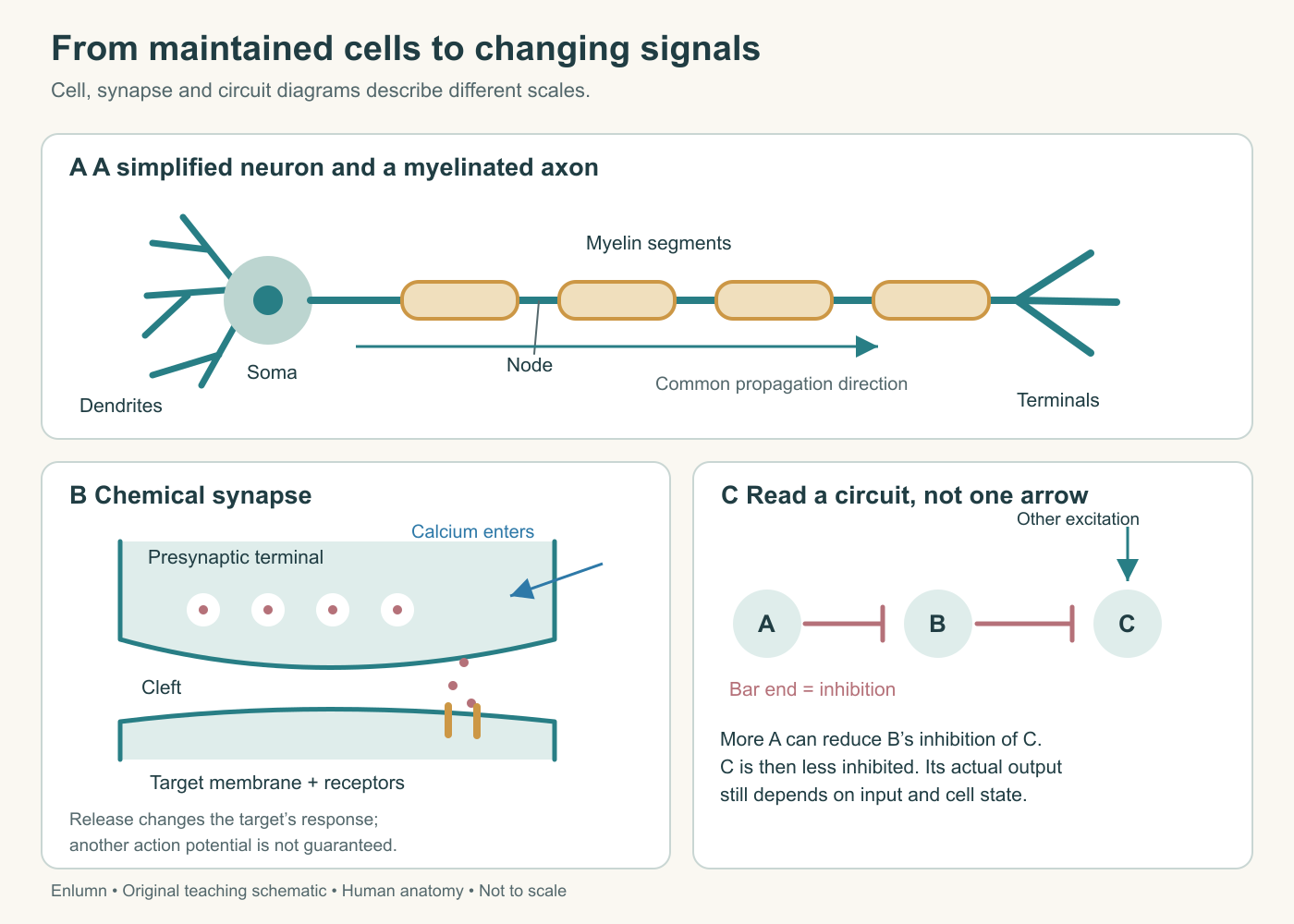
The three panels change scale: a cell, a connection between cells, and a small circuit. White circles in the terminal represent transmitter-containing vesicles; small pink dots represent released transmitter. The lower-right circuit includes independent excitation to C because removing inhibition alone does not guarantee a new action potential.
Crossing a chemical synapse
At a typical chemical synapse, an arriving action potential opens voltage-sensitive calcium channels in the terminal. Calcium enters from outside and helps trigger vesicle fusion. Released neurotransmitter crosses the synaptic cleft and binds receptors on the receiving cell. This sequence converts an electrical change into chemical communication and then into changes in the target cell. It need not produce another action potential. Purves and colleagues describe the sequence in their account of chemical synapses.
Consider the importance of the calcium step. If the action potential reaches the terminal but appropriate transmitter release fails, an upstream recording can look normal while communication across the synapse changes. If release occurs but the receiving receptors respond differently, the same presynaptic event can have a different downstream effect. These hypothetical failures separate propagation, secretion, and reception. They also show why one electrical recording is not a complete account of a connection.
A receptor is a protein with particular molecular interactions. An ionotropic receptor combines transmitter recognition with an ion-channel function. A metabotropic receptor influences intracellular signaling through intermediary mechanisms, often producing slower or longer-lasting effects. Neither category is simply “stronger.” A fast response and a prolonged change in responsiveness solve different problems, and both can affect how subsequent input is processed.
The effect depends on the receptor, the ion channels involved, and the receiving cell's state. Calling a transmitter a chemical for happiness, attention, or courage skips these mechanisms and assigns a complex human description to a molecule. A transmitter can act at multiple receptor types in multiple circuits. Its contribution to behavior cannot be inferred from its name alone. The reference on postsynaptic receptor actions makes the dependence on receptor and cellular machinery explicit.
After release, transmitter must stop acting on the same receptors indefinitely. Depending on the transmitter and synapse, diffusion, uptake into cells, and enzymatic breakdown contribute to ending or reshaping its effect. This is part of communication, not simply cleanup after communication. A message whose effect persists longer changes the timing of the next interaction. Release and removal together help determine what the target experiences.
Combining inputs without a tiny decision maker
An excitatory influence increases the likelihood of a postsynaptic action potential under the conditions being considered. An inhibitory influence decreases it. These terms describe effects on activity, not whether the resulting behavior is beneficial or harmful. An inhibitory neuron can contribute to a useful action by limiting competing activity. More excitation throughout a network does not automatically mean better thinking or stronger performance.
Spatial summation concerns effects of inputs arriving at different locations. Temporal summation concerns effects overlapping in time. To see why timing matters, use an intentionally simplified model. Let a fictional cell begin at negative seventy units of voltage, with a threshold at negative fifty-five. Let each input contribute a five-unit depolarizing change at the site where the model evaluates threshold. If three changes overlap fully, their total reaches the model threshold. If each decays before the next arrives, none reaches it.
The arithmetic is useful because it makes the stated assumptions visible. Real voltage changes do not always add linearly. Their size at the axon's initiation region depends on location, membrane properties, and interactions with other conductances. A five-unit change at a remote dendritic site cannot simply be assumed to remain five units everywhere. Our example demonstrates overlap; it does not estimate a human neuron's true threshold or the number of synapses needed to fire it.
Now add an inhibitory conductance. One possible effect is to make the membrane more negative. Another is to make a simultaneous excitatory input less effective without a large negative shift. This latter influence is often called shunting inhibition: the changed membrane conductance reduces the impact of other input. It is why inhibition should not be defined solely as drawing a downward voltage arrow. The functional question is whether the cell becomes less likely to generate the relevant output.
There is no little observer inside the soma counting votes. Integration is the outcome of physical processes in a cell with a particular geometry and history. “Decision” can be a convenient metaphor, but the mechanism consists of channels, currents, membrane properties, and their interactions. Keeping the mechanism visible avoids explaining neural behavior by inserting a miniature version of the person we are trying to understand.
A circuit changes the meaning of an arrow
Draw three model neurons: A inhibits B, and B inhibits C. Assume B is active enough to suppress C and that C receives some ongoing excitation from elsewhere. Increasing A's inhibitory effect can reduce B's activity. With less inhibition from B, C may become more active. This is disinhibition: reducing an inhibitory influence can increase the target's activity.
The additional excitation to C is an essential part of this example. Removing a brake does not by itself guarantee movement if nothing drives the system. A diagram that omits all background input should therefore say that C becomes less inhibited, rather than promising that C must fire. This small distinction makes a circuit explanation more precise and will help when Chapter 5 discusses movement selection.
A second model has A exciting B, while B excites an inhibitory cell D that feeds back onto B. The initial input can increase B's activity, but the feedback route can subsequently limit it. Depending on strengths and delays, the circuit's time course can differ from that of an isolated excitatory connection. We have not derived an actual brain rhythm; we have shown why a single arrow's sign is insufficient to predict a network's behavior.
Feedforward and feedback describe relationships within a chosen circuit model. A feedforward route carries influence toward a later processing stage; a feedback route returns influence toward an earlier stage. The labels are relative to the pathway being described. Real brain networks contain many loops, so the distinction cannot be read from front and back on an anatomical drawing. A posterior area can send feedback to an anterior one, or vice versa, depending on the processing relationship.
Some neurons also communicate through electrical synapses, where gap junctions permit direct current flow between connected cells. These differ from vesicle-mediated chemical transmission and can support rapid coordination. They are part of the human nervous system, though chemical synapses dominate most introductory circuit accounts. The electrical-synapse reference provides the structural distinction. Our model does not assume that all connections have the same directionality, delay, or molecular mechanism.
What the recording does not say
Suppose a researcher records more firing in a group of cells when someone reaches for a cup than when the person sits still. The observation establishes a difference under those task conditions. It does not by itself reveal whether the cells represent the cup, prepare the movement, process touch, track attention, or contribute to several of those processes. The task changed more than one thing. A better experiment introduces comparisons that separate plausible explanations.
For instance, compare looking at the cup without reaching, reaching to a visible mark without grasping, and grasping a cup after its location has been specified. Each comparison changes the questions the data can address. None is magically pure: even looking without moving involves eye movements and attention. Experimental design tries to make competing interpretations distinguishable rather than to create a person with only one process operating at a time.
At the cellular scale, the same caution applies. A rise in a recorded signal is not automatically increased output from every nearby neuron. The answer depends on what the instrument measures. A membrane recording, an extracellular electrode, and a blood-sensitive image sample different physical events. Chapter 8 will return to measurement; for now, keep the chain separate: the physical event, the recording, the task comparison, and the proposed interpretation.
This leaves us with a workable account of communication. Cells maintain gradients; channels generate changing currents; axons propagate events; synapses alter target cells; circuits transform the effects of individual connections. Seeing the cup and reaching for it depends on organized activity across those mechanisms. The next question is how sensory input enters that organization and becomes useful information about the world.
Check your understanding: In the A-inhibits-B-inhibits-C circuit, why can increasing A's activity make C more active, and why is that increase not guaranteed?
Expected answer: A can reduce B's inhibition of C, producing disinhibition. C becomes more likely to respond to other input, but its actual activity still depends on that input and its cellular state. Removing inhibition is not equivalent to supplying excitation.
Application
Allow 15–20 minutes. Draw the three-cell disinhibition circuit and add an independent excitatory input to C. Label every connection by its effect rather than using an unexplained arrow. Predict what happens when A increases, when the independent input disappears, and when the B-to-C connection is removed.
Then reconstruct the two recording-site calculation. Show the distance conversion, time conversion, and inferred speed. Finish with two sentences explaining why a failed synapse can coexist with a normal upstream action potential. Your answer should distinguish propagation from release and reception, without treating a spike as neurotransmitter traveling down an axon.