The Retina Changes the Kind of Signal
Suppose an optical system forms an excellent image on a sheet of transparent plastic. The sheet will not tell a brain that a door is opening. Focusing has arranged light in space, but it has not supplied the cellular machinery for detecting changes, comparing neighboring regions, or transmitting information. The retina performs these biological tasks while remaining part of the optical path.
The central question is not how a picture is squeezed into a nerve. It is how absorbed light changes a cell, how that change influences other cells, and what features of the incoming pattern survive in neural output. The answer requires several levels of explanation: molecules, membrane voltage, synapses, circuits, and a population of output fibers.
A cell is not the same as one segment of that cell
A rod or cone has a specialized outer segment containing the light-sensitive machinery, an inner segment supporting cellular metabolism, a cell body containing its nucleus, and a synaptic terminal. These are parts of a neuron. A retinal section may place them in different named layers, but the layer boundaries do not turn each part into a separate cell.
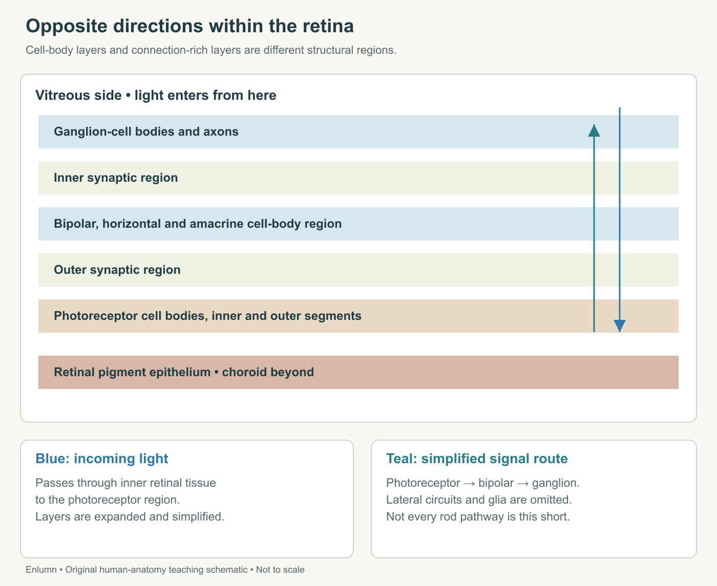
Photoreceptor outer segments lie toward the retinal pigment epithelium, away from the vitreous. Cell bodies occupy the outer nuclear layer, while their terminals participate in the outer plexiform layer. “Plexiform” identifies a region rich in neural processes and connections. “Nuclear” identifies a region containing many cell bodies. These names describe organization, not successive steps that a photon must activate one by one.
Bipolar cells link photoreceptor signals to inner retinal circuitry. Horizontal cells contribute interactions across the outer retina, and amacrine cells contribute diverse interactions in the inner retina. Ganglion cells produce the output whose axons gather into the optic nerve. The retinal anatomy account introduces these cell classes and their lateral as well as forward connections.
A tissue stain that highlights nuclei can make connection-rich layers look relatively empty. That appearance does not mean nothing happens there: many synapses lie among processes extending away from cell bodies.
A minimum circuit drawn as photoreceptor → bipolar cell → ganglion cell is useful, especially for selected cone pathways. It is incomplete. Rod pathways can involve additional interneurons, and many retinal computations depend on lateral interactions. Do not promote a short teaching route into a claim that every signal crosses exactly the same number of synapses.
Absorption starts a biochemical change
Phototransduction is the conversion of absorbed light into a cellular electrical response. Visual pigments contain an opsin protein associated with retinal, a light-absorbing molecule related to vitamin A. Here “retinal” names a molecule; “retina” names the tissue. The similar words are a common source of confusion.
In a rod, photon absorption changes the configuration of retinal within rhodopsin. Activated pigment initiates a cascade involving the G protein transducin and a phosphodiesterase. The cascade reduces cyclic GMP, or cGMP, a signaling molecule that influences channels in the outer-segment membrane. Fewer of these channels remain open, reducing inward positive-ion current and making the membrane potential more negative.
This more-negative shift is hyperpolarization. It reduces glutamate release at the photoreceptor terminal. The phototransduction chapter describes the sequence from pigment activation to changed transmitter release. For the present purpose, retain its direction: light absorption can lead to less transmitter, not necessarily more. Cone transduction follows related principles, with differences in response properties.
The photon supplies the initiating event, but the electrical response depends on a cell maintaining ion gradients and biochemical machinery. A photoreceptor is not a solar panel that simply sends harvested light energy down a wire. Metabolism supports the system in both darkness and light. A detector can consume energy while monitoring an environment even when its preferred stimulus is absent.
Darkness is an active baseline
Rods and cones maintain relatively depolarized states in darkness compared with their light responses. Their terminals release transmitter at baseline. Light changes that ongoing activity. Describing darkness as “the photoreceptor is switched off” misses the actual relationship between stimulus and signal.
Consider a fictional transmitter scale. A receptor releases 12 units during one reference condition and 5 during a brighter condition. The decrease of 7 units carries information because downstream cells respond to changes in release. Nothing requires the meaningful signal to be an increase. A thermostat can convey a change by opening a contact or closing it, provided the receiving circuit is organized accordingly.
The analogy should stop before equating a photoreceptor with a two-state switch. Its membrane response is graded over a range and changes with adaptation and timing. Under ordinary conditions, rods and cones communicate through graded potentials rather than sending conventional trains of action potentials from their outer segments to the brain.
Ganglion-cell axons do carry action potentials. Their timing and rate contribute to neural coding, but a brighter stimulus does not simply produce a proportionally taller action potential. The size of an individual spike and the pattern of spikes are different variables. A fictional trace showing five spikes instead of two represents changed activity without requiring each spike to become two and a half times taller.
The receiving receptor determines the sign
Glutamate is not a command with the same effect at every synapse. Its effect depends on the receptors and cellular mechanisms of the receiving cell. Selected OFF bipolar pathways preserve the sign of the photoreceptor's response: reduced photoreceptor glutamate during a light increment tends to reduce their depolarizing input. Selected ON bipolar pathways invert that relationship, so a light increment can depolarize them.
“ON” and “OFF” refer to response organization, not whether the cell is alive or available for use. Both populations can have ongoing activity and dynamic responses. The account of retinal luminance circuits connects these pathways to detecting increments and decrements in light. The basic lesson is that the same presynaptic change can support different downstream messages.
Use a fictional rule system to isolate the logic. Receiver A has output equal to the transmitter input. Receiver B has output equal to 20 minus that input. When input falls from 12 to 5, A falls from 12 to 5 while B rises from 8 to 15. Opposite outputs have emerged from one input change because the receiving rules differ.
These equations are not models of actual receptor kinetics. They deliberately remove most biology so that sign inversion is visible. Real cells have thresholds, saturation, multiple inputs, and time-dependent behavior. The equations establish a logical possibility; the anatomy and physiology establish that sign differences are used in retinal circuits.
A receptive field is a relationship, not a visible circle
A neuron's receptive field is the region of visual space over which stimulation can influence its response under specified conditions. It is mapped from responses, not necessarily visible as a circular border in the tissue. Neighboring neurons can have overlapping fields. One location in an image can therefore influence several outputs.
Many retinal circuits compare a central region with surrounding regions. In a simplified ON-center arrangement, a light increment in the center increases output while an increment in the surround opposes that effect. OFF-center arrangements reverse the main contrast preference. The retina also contains other response types; these two diagrams do not inventory every ganglion cell.
An original numerical model makes the comparison explicit. Define response drive as center illumination minus average surround illumination, with both measured in arbitrary units. A center of 8 and surround of 3 gives a drive of 5. A center of 8 and surround of 8 gives zero. The center itself has not changed, but its relationship to the surroundings has.
Now add 10 units to both center and surround. The first pattern becomes 18 minus 13, still 5. In this ideal difference model, a common increase cancels. Actual retinal responses are more complex and adaptive, but comparison can emphasize boundaries while reducing sensitivity to a uniform component of illumination.
Do not interpret zero drive as a dead cell or absent vision. It means the two terms cancel in this particular invented calculation. A real cell can retain baseline activity, and other cell populations can report other aspects of the scene. The chosen model describes one operation rather than the entire contents of visual experience.
The lateral connections explain why copying each receptor's value into an isolated output channel would lose part of the retina's work. A circuit can represent how a small region differs from its surroundings rather than merely reporting the absolute amount of light at one point.
Spatial pattern becomes a pattern across cells
Imagine four model output cells with overlapping receptive fields arranged along a dark-to-light boundary. A cell whose center and surround both lie on the dark side may have little difference drive. Another whose center lies on the light side while much of its surround lies on the dark side may respond strongly. Shifting the boundary changes which cells have the stronger response.
The output is thus a distributed pattern tied to spatial relationships. No single cell needs to announce “door edge at this exact coordinate.” Later circuits combine and transform signals from many cells. A neuron can be informative about a feature without uniquely identifying an object or containing the whole percept.
Timing adds another dimension. Some responses emphasize sustained conditions, others changes over time. A moving edge can generate a sequence across cells even if the total amount of light in the scene stays similar. Counting total spikes across every output and discarding their identities and timing would remove useful information.
For an original example, compare two sequences from cells A and B: A responds first and B second, versus B first and A second. Each sequence contains the same number of events. Their order differs, potentially distinguishing opposite movement directions when embedded in an appropriate circuit. A total count of two cannot preserve that difference.
Resetting is part of detecting
After activation, transduction components must return toward states that permit further responses. Signaling cascades are limited and photopigment components are recycled. Outer-segment material is renewed through interaction with the retinal pigment epithelium. Detection therefore depends on maintenance over time, not just the availability of a light-sensitive molecule at one instant.
If an invented detector responds strongly to the first event but never recovers, it cannot distinguish a second event from a continuous first one. A larger first response would not solve that problem. Useful signaling requires a relation between activation, recovery, and the time scale of the environment being monitored.
Adaptation also changes sensitivity as background conditions change. A response measured after one light history need not match a response measured after another. When comparing two records, ask about preceding illumination, test timing, and the measurement itself. “Same light” can mean the same current stimulus while omitting a different state of the receiving system.
The next chapter develops this point for detail, color, and dim scenes. Here it completes the cellular explanation: the retina transforms an input through machinery whose state depends partly on earlier inputs. It is neither a blank photographic plate nor a fixed mathematical filter.
Image formation is not the eye's only use of light
A subset of ganglion cells contains melanopsin and can respond to light intrinsically, in addition to receiving retinal input. These cells contribute to functions including pupil regulation and alignment of circadian timing with environmental light. The review by Markwell and colleagues describes these contributions. “Ganglion cell” therefore does not mean every such cell is only a passive recipient of rod and cone signals.
Zaidi and colleagues reported selected light responses in two profoundly blind participants lacking functional rods and cones. The findings included circadian or pupillary responses under particular conditions and limited light awareness in one participant. They show why absence of conventional image-forming vision does not necessarily mean absence of every biological response to light.
Two cases do not establish the same retained function in every blind person. Nor do they prescribe a light exposure or prove that melanopsin can recreate normal detailed vision. The study is useful because it separates outcomes that everyday language can collapse into the single word “seeing.”
The retina changes the kind of signal while preserving and transforming useful relationships. Photons are absorbed, molecules change state, voltage and transmitter release change, circuits compare inputs, and axons carry structured output. Each step offers a distinct explanation for what the next step receives.
Check your understanding: Light reduces transmitter release from a photoreceptor. Must every downstream cell reduce its activity, and does equal total ganglion-cell spike count establish identical visual information?
Expected answer: No. Different postsynaptic mechanisms can preserve or invert the effect of changed transmitter release. Equal total spike counts also omit which cells responded and when; different spatial and temporal patterns can carry different information.
Application
Allow fifteen to twenty minutes. Draw two arrows through a retinal section: incoming light toward photoreceptor outer segments and selected neural signaling toward ganglion-cell output. Include the retinal pigment epithelium, photoreceptor cell body and terminal, bipolar cell, one lateral interaction and a ganglion-cell axon.
For the supplied difference model, calculate drive for center/surround pairs 12/4, 12/12 and 22/14. Then explain why equal drive in two cases does not prove identical human perception. Use the fictional receiver rules A = input and B = 20 − input to calculate outputs when transmitter input changes from 9 to 6.
A strong response obtains drives 8, 0 and 8; A changes from 9 to 6 while B changes from 11 to 14. It labels the equations as limited teaching models, distinguishes graded receptor signaling from axonal spikes, and avoids treating the human light-response cases as a universal claim about blindness.