Where vision can be disrupted
Three fictional eyes receive light from the same striped sign. In the first, the stripes are projected out of focus onto a responsive retina. In the second, the lens scatters light even though its average focusing power is appropriate. In the third, a clear, focused image reaches tissue that cannot produce the expected neural output. The reports might all include the word “blur.” Yet the word does not identify which physical process has changed. We can make more progress by asking where the information becomes less useful, what a proposed correction actually changes, and what observations would distinguish competing explanations.
Follow two routes rather than one arrow
The light route runs through the tear-covered cornea, aqueous humor, pupil, lens, and vitreous toward the photoreceptors. Along it, surfaces redirect light, the aperture restricts entry, and transparent tissues must remain sufficiently clear. At the receptors, absorbed light initiates changes in cellular signaling. The useful connection between these routes is transduction. A photon does not become an action potential that travels intact through a hollow optic nerve.
For the major pathway supporting vision, retinal ganglion-cell axons enter the optic nerve, undergo partial crossing at the optic chiasm, and continue in optic tracts. Many terminate in the lateral geniculate nucleus of the thalamus. Neurons there project through the optic radiations to primary visual cortex in the occipital lobe. The tract on either side therefore contains fibers from both eyes. Other retinal projections participate in pupil regulation, orienting movements, and daily rhythms. This is a branching nervous system, not a single cable ending at a screen. Purves and colleagues, Central Projections of Retinal Ganglion Cells.
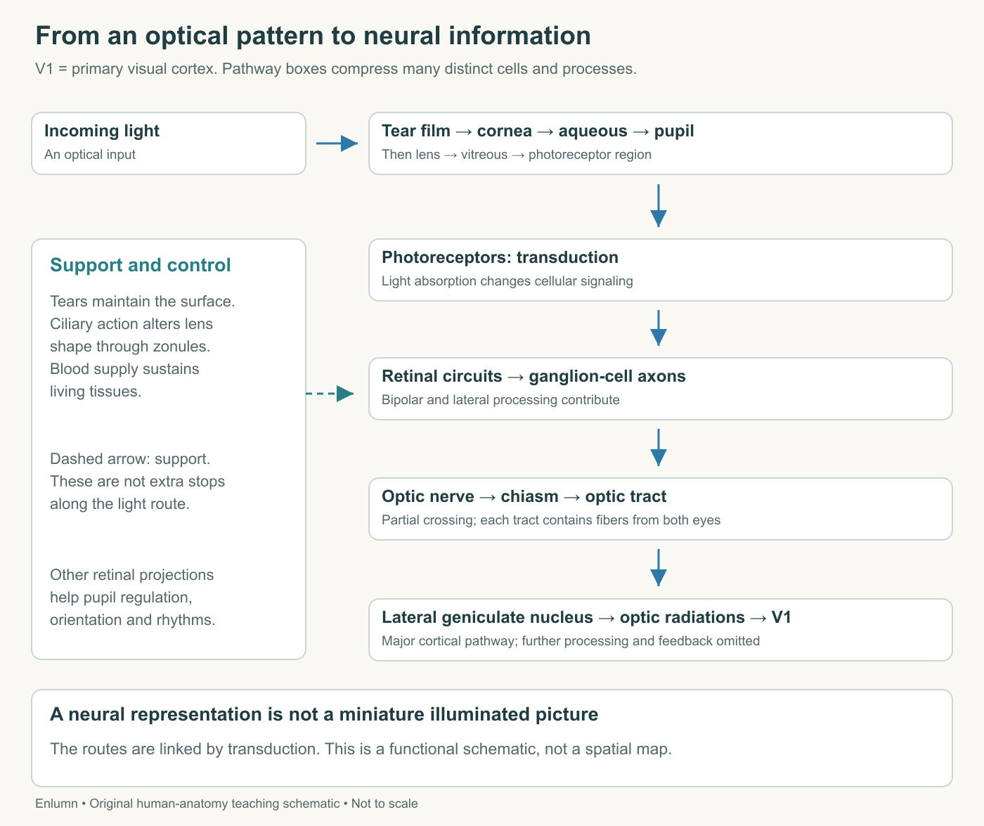
The diagram deliberately leaves out most synapses and feedback connections. Its arrows label relationships, not anatomical distances or measured transmission times. Notice especially that the ciliary body belongs in a support and control relationship to the optical system. Adding it as a compulsory stop after the lens would misrepresent the route of incoming light. Likewise, blood vessels supply living tissue; they do not carry a photographic image from retina to cortex. Diagram errors often begin when different kinds of arrow are given the same meaning.
A focusing problem is a geometrical problem
Return to fictional eye A. Its optical model forms a sharp image on a plane slightly in front of the retina. On the retinal plane, light from one object point has already spread into a small patch. Two nearby points may produce overlapping patches. A change in effective optical power could bring those patches together at the retinal surface, improving the distinction between them without adding photoreceptors or replacing neurons.
Suppose a simplified demonstration has two bright points separated by four arbitrary distance units. Each produces a patch six units wide at the screen. The patches overlap. After adjusting focus, each patch is one unit wide, while their centers remain four units apart. The demonstration predicts better separation. It does not predict a particular person's visual acuity: actual images contain diffraction, aberrations, scatter, varied contrast, retinal sampling, and processing beyond the eye. The numerical patches isolate one relationship so we can reason about it.
The strongest evidence for the model would combine a measured change in image geometry with improved discrimination under otherwise comparable conditions. Merely reporting “the lens was changed” does not establish that the intended correction occurred. A changed lens might also alter magnification or transmitted light. In an experiment, those possibilities become variables to measure or control. This is the difference between naming an intervention and identifying its mechanism.
Refractive conditions also need their qualifications. Myopia places the relaxed eye's focus for distant light in front of the retina; hyperopia places it behind. Accommodation may compensate for some hyperopic defocus. Astigmatism involves differing optical power with orientation. Presbyopia concerns reduced accommodation with age. These descriptions can overlap within one person. They are not four mutually exclusive kinds of eye. National Eye Institute, Types of Refractive Errors.
Clarity can fail even when average focus is appropriate
Eye B poses a different problem. Imagine that the intended bright stripe delivers 90 units to its location and the dark stripe receives 10. Define a contrast measure for this exercise as the difference divided by the sum: (90 − 10)/(90 + 10) = 0.8. Now redistribute some light so the bright location receives 70 and the dark one 30. The sum is still 100, but the contrast is 0.4. The demonstration has not simply dimmed the image. It has reduced the distinction between neighboring regions.
Compare a neutral reduction in light: values of 45 and 5 also total less light, but their contrast is (45 − 5)/(45 + 5) = 0.8. This does not make reduced illumination harmless; photon availability and neural noise still matter. It shows why total transmitted light alone cannot describe image quality. Two systems delivering the same total can distribute it differently, and two delivering different totals can preserve the same normalized contrast.
A cataract is clouding within the lens. It can interfere with clear vision and contribute to glare or altered color appearance. A corrective spectacle lens changes optical power; it does not remove clouded lens tissue. Replacing the clouded lens addresses that optical component, while the result also depends on the rest of the visual system. The condition is not a film growing over the outside of the eye, nor does every blurred image establish its presence. National Eye Institute, Cataracts.
Our contrast exercise is an invented redistribution model, not a measured cataract profile. It omits wavelength dependence, angular scattering, and the complex light distribution across a scene. Its strength is narrower: it distinguishes sharpening a defocused image from preserving differences when unwanted light reaches a nominally dark region. If someone claims that increasing brightness necessarily fixes both, the model provides a counterexample. Multiplying 70 and 30 by two leaves the normalized contrast at 0.4.
A receiving surface must remain living and connected
In eye C, the optics deliver distinct patterns, but the expected neural responses do not follow. A useful model separates the input image from the tissue's capacity to use it. Suppose five sampled positions receive values 2, 8, 2, 8, and 2. An intact teaching model reports those five values. If two positions supply no usable report, the available sequence might be 2, missing, 2, missing, 2. Calling the missing reports zero would insert information that was never measured.
That distinction matters. Zero could mean a functioning system measured darkness; missing could mean the relevant signal could not be obtained. The brain does not literally receive a spreadsheet containing the word “missing,” and visual experience is not a direct reading of our table. We use the table to expose a reasoning mistake: an absent or altered neural report cannot be repaired simply by assuming that the corresponding region of the world was dark.
Retinal detachment separates retina from its normal position against the back of the eye and threatens its function. Its mechanisms include more than a single universal story of a tear. An important real-world boundary belongs here: sudden new floaters, flashes, or a curtain-like shadow can signal an emergency requiring immediate eye or emergency care. The fictional tables in this course cannot decide what an individual's symptoms mean. National Eye Institute, Retinal Detachment.
A neural problem can also occur after local retinal processing. The optic nerve consists of axons, not transparent optical material. Changing corneal refraction cannot recreate a lost axon. Conversely, evidence that the optic nerve is structurally present does not prove that every upstream receptor or downstream circuit functions normally. Anatomy constrains an explanation, but a picture of a structure and a measurement of its performance answer different questions.
Separate a risk indicator from the outcome of interest
Eye pressure illustrates another common inferential error. Elevated pressure increases glaucoma risk, but some people with elevated pressure do not develop glaucoma, and glaucoma can occur without pressure above the usual range. A pressure value is therefore not interchangeable with evidence about optic-nerve damage or visual function. National Eye Institute, Eye Pressure.
Consider a completely fictional device study rather than a clinical dataset. Of 100 devices exposed to high mechanical load, 30 later lose signal. Of 100 at lower load, 10 lose signal. The high-load group's failure proportion is three times as large, yet 70 high-load devices do not fail and 10 lower-load devices do. The association identifies a risk difference without producing a perfect classification rule. These numbers are invented and tell us nothing about human disease rates.
Even that device association is not automatically causal. Perhaps the higher-load devices are older, operate at higher temperatures, or receive less maintenance. A well-designed investigation asks how groups were formed and which other differences might explain the result. It also defines failure consistently. If one laboratory calls a small decline a failure and another requires complete loss, their percentages cannot be compared as though they measured the same endpoint.
Now imagine an intervention lowers the recorded load. That is evidence about the load measurement. To establish improved performance, investigators must also assess the relevant functional outcome over an appropriate interval. The example introduces a general rule for reading evidence: specify which link was measured. A successful change in a proposed cause is encouraging, but the desired consequence remains a separate empirical question.
Build a comparison that can actually teach you something
Here is a supplied test bench for the three fictional eyes. An optical instrument measures the retinal-plane pattern; a separate sensor records neural output. A final behavioral task asks the modeled observer to distinguish stripe orientations. In A, a focusing adjustment improves the retinal-plane pattern and the orientation result. In B, adjustment changes the focus setting but leaves the scattered-light contrast loss. In C, the optical pattern remains distinct while the neural response is impaired. These are stipulated teaching observations, not diagnostic instructions.
The three measurements occupy different positions in an explanatory chain. If the task improves while the optical measurement does not, you should not silently announce an optical mechanism. Learning, attention, measurement sensitivity, or a different unmeasured change might matter. If the optical pattern improves while task performance does not, that also deserves explanation. The task might demand distinctions beyond available sampling, or another process might limit performance. A result can challenge the proposed mechanism without making either instrument meaningless.
Add one further complication: two impairments can coexist. A model with defocus and reduced neural output may improve after optical correction without reaching the performance of a model with neither limitation. Partial improvement is compatible with correcting a real component. It does not show that the remaining limitation is imaginary, and it does not prove which remaining component matters. This is why comparisons need both a proposed mechanism and an explicit boundary around the conclusion.
What the completed model should allow you to explain
A strong account can answer more than “what is this part called?” It can predict why reducing pupil diameter changes light entry without replacing photoreceptors; why increasing optical power changes focus without making a cloudy lens transparent; and why a responsive pupil alone does not describe every process required for detailed vision. Each answer identifies an input, a transformation, and a consequence. It also states what the observation leaves unresolved.
Consider a museum display that labels the eye “a camera that sends pictures to the brain.” The analogy helps with aperture and focusing surfaces, but its final phrase hides the most consequential transformation. Retinal circuits generate patterns of neural activity rather than transmitting a miniature illuminated scene. Supporting tissues continually sustain the apparatus. Your improved label should retain the useful optical comparison while explaining the living conversion and avoiding the idea of an internal viewer examining a screen.
Check your understanding: In the fictional test bench, an optical adjustment sharpens the retinal-plane pattern but does not improve the orientation task. What has been demonstrated, and what remains unresolved?
Expected answer: The adjustment improved the measured optical pattern. It has not established improved task performance or identified its remaining limitation. Retinal sampling, neural function, task conditions, or measurement limits may require investigation; the result does not show that optical correction was physically ineffective.
Application
Allow 30–40 minutes. Produce two linked diagrams and a 600–900-word explanation for a learner who has not taken this course. The first traces incoming light through the eye. The second begins with photoreceptor signaling, includes retinal processing and ganglion-cell output, and reaches the major brain pathway. Use a separate arrow style for blood supply, tear-film maintenance, and ciliary control. State that the diagrams omit most cellular detail and central feedback.
Explain three supplied disruptions: A forms its best image in front of the retinal plane; B redistributes a 90/10 stripe pattern into 70/30 while preserving total light; C receives a distinct pattern but has impaired ganglion-cell output. For each, identify the changed process, calculate anything supported by the data, and explain why one intervention directed at another process would not directly repair it. Then add a fourth case with both A and C and explain possible partial improvement.
Finish by revising the museum label in fewer than 70 words. Check your work against four criteria: light and neural activity have different routes; named structures have specific roles; the contrast calculation yields 0.8 and 0.4; and none of the cases becomes a rule for diagnosing a real person's vision. A successful explanation separates optical formation, living transduction, and perception while showing how they depend on one another.