Adjusting water and salts
A kidney can change the amount of water leaving with a given solute load. That capacity matters because the body does not receive water and dissolved substances in a perfectly fixed ratio. Food, drinking, metabolism and losses create changing conditions. A useful urinary system must adjust its output while continuing to remove material that cannot simply accumulate indefinitely.
The adjustment depends on more than a command to “save water.” It requires a gradient that can drive water movement, an epithelial route whose permeability can change, and signals that connect the kidney to conditions elsewhere. This chapter assembles those three parts before using them in a supplied water-balance case.
Amount and concentration are different control problems
Start with a sealed model compartment containing 100 solute units in ten water-volume units. Its concentration is ten. Remove two water-volume units while leaving all the solute: concentration becomes 12.5. Instead, remove 20 solute units and two water-volume units together: concentration remains ten, although the total contents are smaller.
The first change alters concentration strongly; the second changes volume without changing the ratio. These are not equivalent disturbances, even if the same amount of water leaves. A regulatory system concerned with concentration needs information different from a system concerned with effective circulating volume.
Physiology often expresses the total concentration of dissolved particles as osmolality, the number of osmotically active particles per mass of water. Osmolarity uses solution volume instead. For this introductory course, the central idea is that water balance changes the relation between dissolved particles and their solvent. Our invented “solute units per water-volume unit” examples teach that relation without serving as laboratory reference values.
A sodium concentration is also a ratio, not a direct inventory of all sodium in the body. The same measured ratio can accompany different total amounts and distributions of water and solute. That is one reason a single concentration cannot stand in for an entire fluid-balance assessment.
Water needs a pathway and a driving difference
Place two model chambers beside each other, separated by a wall that passes water but retains the stipulated solute. If their effective osmotic conditions differ, net water movement can occur toward the side with the greater retained-solute concentration, until opposing influences balance it. If the wall is effectively impermeable to water, the concentration difference alone produces little transfer over the interval considered.
Now make the wall highly water-permeable but remove the concentration difference. Opening more pathways does not manufacture a directional osmotic driving force. Permeability and gradient answer different questions: how readily can water cross, and which influences favor net movement?
Keep those questions together when reading about aquaporins. They are water channels, not pumps that spend energy to push water uphill. Changing the number of available channels changes the ease of crossing. Other processes establish and maintain the conditions under which that crossing results in net recovery.
The kidney's concentrating apparatus combines tubular transport, the medullary environment, circulation and hormonal regulation. Sodium chloride and urea contribute substantially to the high solute concentration of the medullary interstitium, with urea particularly important in the inner medulla. Agaba and colleagues: concentrating mechanism review, indexed text.
Different loop segments do different work
In a simplified loop model, water-permeable descending regions permit water to leave as fluid travels into a more concentrated environment. The thick ascending limb reabsorbs salt while having very low water permeability, allowing tubular fluid to become more dilute as it moves upward. The real loop contains several segments; their properties should not be collapsed into one uniform wall. OpenStax: loop segments.
There is an important consequence: fluid can become concentrated at one point along a nephron and more dilute farther along. The direction of the route does not require its concentration to rise steadily. A graph of tubular concentration against position can bend because the transport properties change with position.
For a model thick ascending segment, begin with ten solute units in ten water-volume units. Remove six solute units while retaining all ten water units. The outgoing concentration becomes 0.4 rather than one. The surrounding compartment has gained solute, while the tubular compartment has become more dilute. Both outcomes follow from the same transfer.
Now place a water-permeable segment nearby, with fluid traveling in the opposite direction. It can respond to the local environment created in part by salt transport. With continuing flow and repeated effects along the route, a small local difference can contribute to a larger difference along the medulla. This is the logic of countercurrent multiplication.
The loop is not creating solute from nothing or extracting unlimited water. Energy-dependent transport, incoming material, geometry and flow support the gradient. Remove those supports and the behavior changes. A U shape alone does not produce concentration; it must be paired with appropriate transport properties.
The surrounding blood flow helps preserve the arrangement
The vasa recta carry blood through the medullary environment and exchange water and solutes along their course. Their countercurrent organization limits rapid removal of medullary solutes while permitting recovered water to return to circulation. This is countercurrent exchange, distinct from the transport processes that establish the gradient. Agaba and colleagues: medullary circulation.
An original two-level thought experiment helps. Suppose blood descending into a concentrated region gains solute. On its return toward a less concentrated region, some of that solute can leave again. The arrangement reduces how much is carried all the way out compared with an otherwise similar route that simply enters the concentrated region and departs without a return exchange path.
This is a qualitative explanation, not a complete vascular simulation. Real vessels branch, occupy particular spatial relationships and carry oxygen as well as exchange material. The useful distinction is between maintaining local concentration and supplying a living tissue; the circulation has to participate in both.
Urea also moves among tubular and interstitial compartments rather than following a single one-way disposal arrow. Recycling helps its contribution to the medullary environment. Detailed accounts of the inner-medullary gradient have involved competing models and unresolved quantitative issues; the basic teaching diagram should not be mistaken for a fully settled reconstruction. Sands and Layton: urinary concentration review.
For this course, retain the experimentally grounded requirements: segment-specific transport, an appropriate medullary environment and regulated water permeability. We do not need to pretend that every microscopic contribution is known exactly to explain why removing any of those requirements can impair concentration.
Vasopressin changes the collecting duct's response
Arginine vasopressin, also called antidiuretic hormone or ADH, is made in hypothalamic neurons and released into circulation from their terminals in the posterior pituitary. Osmotic signals influence its release, and thirst provides a related behavioral response. Vasopressin acts through V2 receptors in the kidney to support collecting-duct water recovery. Endotext: physiology of vasopressin.
AQP2 is an important water channel in this response. Vasopressin signaling promotes its placement in the collecting-duct cell membrane; reduced signaling allows channels to be removed. This changes the water permeability of the luminal membrane and therefore how much water can be recovered under the prevailing conditions. MedlinePlus Genetics: AQP2 function.
The hormone does not reach into finished urine in the bladder and pull water backward through the ureter. It acts on cells along the route while fluid is still being processed. Nor is adding a channel the same event as making a new medullary gradient. The response uses the surrounding conditions as well as modifying the epithelial pathway.
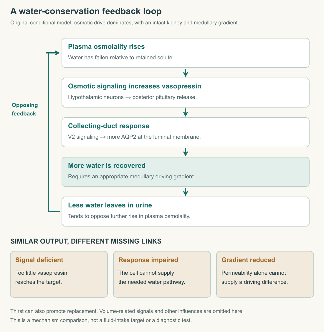
Read the loop all the way back to the starting condition. Increased recovery tends to reduce further water loss, opposing the disturbance. That return influence makes the relation negative feedback. An arrow ending at “more concentrated urine” would describe an output but leave the regulatory purpose unfinished.
Work through a water-loss case without prescribing intake
Consider a stipulated healthy model system in which water loss temporarily exceeds water replacement, while the relevant retained-solute amount changes little. Assume the resulting rise in plasma osmolality is the dominant signal and that the concentrating apparatus can respond. Predict directions of change, not an exact volume or a personal drinking recommendation.
The model predicts increased vasopressin signaling, greater availability of AQP2 at the relevant membrane, increased water recovery and a smaller urinary water loss than would occur without that response. Thirst can also promote replacement. These changes tend to oppose the rise in concentration; they do not instantly erase all prior losses.
Now reverse the supplied disturbance. Water is added in excess of current needs while other stipulated factors remain stable. Lower osmotic drive reduces vasopressin signaling, water recovery through the adjustable route decreases, and more dilute urine can be produced. The response is conditional on intact regulation and excretory capacity.
The word “can” matters. A real person's output depends on more than our two-case model: concurrent solute loads, losses, signaling, medications and disease may change the situation. The exercise teaches a causal loop, not a method for deciding how much someone should drink from the appearance of their urine.
A numerical illustration makes the water-saving effect visible. Suppose 100 water-volume units enter our model tubular route during an interval. Earlier segments recover 85, leaving 15 for a final adjustable stage. Recovering ten there leaves five to exit; recovering 13 leaves two. A three-unit change at the final stage reduces final water output by three-fifths, despite changing only three percent of the initial volume.
This explains why a late adjustment can have a large proportional effect on the final output. A small difference relative to a large upstream stream can be a large difference relative to the small residual stream. The percentages describe different denominators, so both statements can be true.
A response unfolds over time
A feedback diagram is usually drawn without a clock, but the order of events matters. First comes the disturbance. Sensors and signaling respond; cellular behavior changes; the altered output then affects the body's contents over an interval. Drawing these as separate stages prevents the false expectation that a regulatory response must instantly restore the starting condition.
Use an invented four-interval record. A model compartment begins with ten water units. During the first interval, incoming water is one and total loss is two, leaving nine. In the second interval, the same imbalance leaves eight. Suppose conservation then lowers loss to one while incoming water remains one. The third interval ends at eight, and so does the fourth. The response has stopped further decline; it has not replaced the two units already lost.
To restore ten, a later interval must have a net gain. That gain could result from more input, less output or both within the model's constraints. This distinction between stopping a disturbance and reversing its accumulated effect is essential to reading any homeostatic account.
The same arithmetic shows why a signal can remain active after the original external event ends. A past loss may have left the compartment altered, even when its current inflow and outflow match. Feedback responds to the controlled condition, not simply to whether the triggering event is still happening.
For a human explanation, avoid turning these arbitrary units into a timetable or fluid target. Their purpose is to make conservation visible over successive intervals. A full physiological prediction would also track solute, compartment distribution and the changing signals. The simple ledger establishes what any more detailed model must respect: stored amount changes only when the combined inputs and outputs fail to balance.
Sodium and potassium need their own accounting
Aldosterone is released by the adrenal cortex, with angiotensin II and elevated plasma potassium among its stimuli. It promotes sodium reabsorption and potassium secretion in responsive distal nephron regions. Its effects are related to fluid balance but are not identical to vasopressin's regulation of water permeability. OpenStax: aldosterone.
The distinction is easier to see if you draw separate ledgers. One tracks water, one sodium and one potassium. A hormonal signal can change more than one ledger, but they do not become interchangeable. Losing water without proportional solute loss differs from losing sodium with accompanying water, and a potassium change raises another regulatory question.
In an invented distal-segment example, ten sodium units arrive and eight are recovered, leaving two. If recovery rises to nine with delivery fixed, sodium excretion falls to one. Nothing in that arithmetic establishes the exact water change. To predict it, we must also specify water permeability, osmotic conditions and the other substances in the fluid.
Likewise, knowing that a signal can promote potassium secretion does not determine a person's final potassium balance. Delivery to the segment, cellular transport conditions, intake and other routes of loss matter. Hormone names are starting points for a mechanism, not substitutes for all its conditions.
Similar outputs can arise from different failures
A deficit of vasopressin and resistance to its action can both impair water conservation. The former concerns insufficient signal; the latter concerns the kidney's response despite signal availability. These are now also termed AVP deficiency and AVP resistance. Endotext: deficiency and resistance.
AQP2 variants provide a concrete cellular example. Some cause channels to be misfolded or sent to the wrong location, preventing their proper placement at the membrane. A channel protein can therefore be present in a cell without supplying the needed water pathway at its working surface. MedlinePlus Genetics: AQP2-related resistance.
Our gradient model identifies a third possible limitation: a responsive membrane cannot recover water normally if the surrounding driving conditions are disrupted. More signal cannot automatically repair every downstream defect. The causal chain has several necessary links.
For a paper exercise, label three hypothetical systems “signal absent,” “membrane response absent,” and “gradient reduced.” All may produce relatively dilute output under conditions in which the intact model would conserve water. Their shared output does not establish a shared cause. Distinguishing them would require evidence about the relevant signal, response and environment.
Do not turn this comparison into a self-administered water-deprivation experiment. The supplied cases contain the assumptions needed for learning; actual diagnostic testing requires clinical supervision. Here, the useful conclusion is mechanistic: water conservation is a coordinated response across brain, blood, tubular cells and medullary structure.
Application
Draw the feedback loop for the stipulated rise in plasma osmolality. Include the signal, receptor-mediated cellular response, water pathway, change in urinary water loss and the opposing influence on the original disturbance. Add a separate label for the medullary gradient so it does not disappear inside the word “hormone.”
Check your understanding: In the model, 15 water-volume units reach the adjustable stage. Recovery changes from ten to 13. What happens to final output, and why would increasing water-channel availability alone not guarantee the same response if the driving gradient were absent?
Expected answer: Final output falls from five to two volume units, a reduction of three units or 60 percent of the original final output. Channels increase permeability; they do not create the osmotic driving difference. The predicted recovery therefore also depends on the surrounding gradient and other stated conditions.
Write a short comparison of signal deficiency, membrane-response failure and reduced medullary gradient. Identify a different missing piece of evidence for each. A good answer does not diagnose all three from a single dilute sample or equate sodium concentration with total body sodium.