Making substances the body uses
A protein circulating near your ankle may have been assembled in the liver. A bile salt helping with intestinal fat absorption may have left a hepatocyte, completed several rounds through the intestine and returned to be secreted again. Both are liver outputs, but one travels in blood and the other initially travels in bile. To understand production, follow a product beyond the cell that makes it. Its usefulness depends on reaching another compartment, interacting with other molecules and eventually being removed or recycled.
Production begins with cellular work
A hepatocyte making a protein must translate information into an amino-acid sequence, fold and process the resulting molecule, and deliver it to the appropriate destination. Ribosomes, endoplasmic reticulum and Golgi-associated pathways contribute to these steps. Supplying amino acids is necessary but not sufficient. The cell also requires energy, functioning machinery and correctly regulated trafficking.
This gives “synthetic capacity” a more precise meaning. It is the capacity to make particular products at particular rates under given conditions, not simply the mass of an organ. Two equally sized pieces of tissue could differ in the number or state of functioning hepatocytes. A cell that synthesizes a protein but cannot release it is also different from one that never makes it. The location of the failure affects what measurements would reveal.
Imagine a fictional production sequence with four steps: assembly, processing, secretion and survival in circulation. If measured blood levels fall, the problem could arise at any of these steps. A short-lived product may disappear despite normal synthesis; an expanded plasma volume can lower its concentration without changing the amount present. The concentration is an outcome of the whole route, not a direct meter attached to the liver's protein-making machinery.
Albumin connects synthesis to circulation
Albumin is an abundant plasma protein made by hepatocytes. It binds a range of substances, including fatty acids and bilirubin, and contributes strongly to plasma oncotic pressure, the osmotic effect associated with proteins across a selectively permeable vascular interface. The Clinical Methods account of albumin provides a reference for these transport and fluid-distribution roles.
Binding helps explain how poorly water-soluble substances can travel in aqueous blood. The carried substance interacts with sites on albumin; it does not necessarily become permanently attached or chemically converted into albumin. Bound and unbound forms can exchange. This is a transport relationship, distinct from the enzymatic transformations that may occur after the substance reaches a tissue.
For a deliberately simple binding example, imagine 100 sites, each able to hold one unit of a fictional molecule. If 80 units occupy sites, 20 sites remain available under that model's assumptions. Adding 30 more units cannot put one unit on each of the original 100 sites without leaving some unbound. Real binding involves affinity, competing molecules and changing concentrations; the example merely shows why amount and available binding capacity are different questions.
Do not turn that model into a drug-dose rule. Some substances have several binding partners, and their distribution and removal can change together. Even in a classroom diagram, it is better to label the bound pool and the unbound pool separately than to equate total concentration with immediate biological effect. Albumin provides a concrete example of how the physical state of a molecule matters as well as its quantity.
Its role in fluid distribution is equally relational. Water movement across small-vessel walls depends on pressure differences, protein-related osmotic effects, properties of the endothelial barrier and the return of filtered fluid through lymphatic routes. Albumin contributes to that system; it is not a sponge sitting inside a vessel. Lower plasma albumin can favor altered fluid distribution, but it does not by itself explain every instance of swelling or abdominal fluid accumulation.
Compare two invented situations. In one, plasma albumin falls while vascular pressures and permeability initially remain the same. In the other, albumin stays constant but venous pressure rises. Both can change net fluid movement, through different parts of the system. A third situation could involve a more permeable barrier. A useful explanation identifies the changing influence instead of describing all three as the blood “not holding water.”
A circulating pool is not a production rate
Suppose an original model contains 120 units of a protein in a well-defined circulating pool. The liver adds 10 units per day and 10 are removed, so the amount is stable. If removal rises to 15 while synthesis stays at 10, the pool loses 5 per day. After four days under these fixed assumptions, 100 units remain. Synthesis has not fallen, yet the circulating amount has.
Now keep 120 units but increase the fluid volume from 3 to 4 volume units. Concentration falls from 40 to 30 units per volume unit. There has been no loss of the protein in this second model. The two examples produce lower concentrations by different mechanisms. They also show why a concentration measurement cannot independently determine synthesis, distribution and degradation.
Actual albumin occupies both vascular and extravascular spaces, and its turnover is more complex than the single-pool illustration. Changes in inflammation, losses and fluid balance can affect measured levels. The point is not to teach a personal laboratory interpretation but to preserve the distinction between a stock, its concentration and the rates that maintain it. Those distinctions recur throughout organ physiology.
The liver also makes many other circulating proteins. It does not make every plasma protein. Antibodies, for example, are made by plasma cells of the immune system. The OpenStax overview of blood distinguishes albumin, fibrinogen and immunoglobulins. A list of proteins present in blood is not automatically a list of liver products.
Helping clotting means helping its regulation
Fibrinogen is a liver-produced soluble protein that can be converted into fibrin during clot formation. Fibrin forms part of the mesh that stabilizes a clot. The liver also produces many other proteins participating in coagulation and in its control. Platelets, vessel walls, enzymes and local conditions are essential partners. A clot is an organized response at a site, not a lump manufactured in the liver and sent out ready-made.
The distinction between precursor and active product matters. If a circulating precursor can be activated locally, the system can maintain potential capacity without activating it everywhere. A model in which every clotting protein is always fully active would fail to explain how blood usually remains fluid while still being able to form a clot where needed.
Regulation also requires restraints. The liver contributes proteins that limit coagulation as well as proteins that promote it. Consequently, reduced hepatic synthesis cannot be interpreted simply as a uniform reduction of every tendency to clot. The 2021 ISTH communication explains why liver disease can alter opposing parts of hemostasis and produce a fragile balance. The course uses that principle, without giving treatment decisions or inferring an individual's bleeding risk from a routine test.
A thought experiment makes the logic accessible. Imagine a regulated process with an activating influence and a limiting influence. Measuring only the activator misses a simultaneous change in the limit. Reducing both can leave one laboratory output similar while changing its response to a disturbance. This is not a quantitative coagulation model: real hemostasis has many pathways and local interactions. It is a reason to inspect both promoting and restraining mechanisms before predicting the whole-system result.
Bile combines several substances for different purposes
Bile is a mixture of water, ions, bile acids and salts, phospholipids, cholesterol, bilirubin-related material and other constituents. These components do not all do the same job. Some aid intestinal lipid handling, some contribute to fluid properties, and some are being excreted. A fluid can perform a digestive role and carry materials toward elimination at the same time.
Hepatocytes synthesize primary bile acids from cholesterol. Many bile acids are conjugated with glycine or taurine and occur predominantly in ionized forms under physiological conditions. The term bile salts commonly refers to these ionized bile-acid forms. They are amphipathic: their structure includes regions that interact differently with water and with lipids. This property helps explain their work at interfaces and in mixed aggregates.
Consider oil dispersed in water. Dividing a large droplet into smaller droplets increases the available surface at which digestive enzymes can act. That is emulsification, a physical change in dispersion. Hydrolysis, by contrast, breaks particular chemical bonds. Bile salts help the conditions for fat digestion and absorption; they do not replace the lipase enzymes that perform chemical cleavage.
In an original geometry example, a sphere of radius 2 has volume proportional to 8 and area proportional to 4. Eight spheres of radius 1 preserve the same total volume but have total area proportional to 8. The total surface area doubles. The calculation does not describe a measured intestinal droplet size; it shows why changing dispersion can matter even when the amount of lipid remains unchanged.
Bile salts also help form mixed micelles that carry products of lipid digestion through the aqueous environment near the intestinal surface. A micelle is not a chylomicron. The former operates in the intestinal lumen; the latter is a lipoprotein package assembled by intestinal cells for transport after uptake. Nor should a diagram imply that a whole micelle is simply swallowed intact into a villus. Components exchange and are taken up through appropriate routes.
A secretion requires work and a pathway
Bile formation depends on transport at the hepatocyte's canalicular membrane. Moving solutes into the canalicular space helps establish osmotic conditions that draw water into the secretion. Cholangiocytes subsequently modify the fluid through their own absorptive and secretory processes. Boyer's physiological account describes the canalicular and ductal contributions. Bile formation is therefore not simply blood pressure squeezing a filtrate through a passive sieve.
Compare three original scenarios. First, a hepatocyte synthesizes bile acids but cannot export them effectively. Second, canalicular export works but a downstream duct is obstructed. Third, the route is patent but duct-cell fluid modification changes. Each can affect the final secretion, but the first concerns a cellular transport step, the second a route, and the third a downstream epithelial contribution.
The final fluid cannot automatically identify which step changed. If collected bile has a different concentration, one constituent's amount may have changed, water movement may have changed, or both may have changed. This is the same amount-versus-concentration distinction used for albumin, now in another compartment. Reusing the principle is valuable because it lets one reasoning tool explain different physiology without making the two products equivalent.
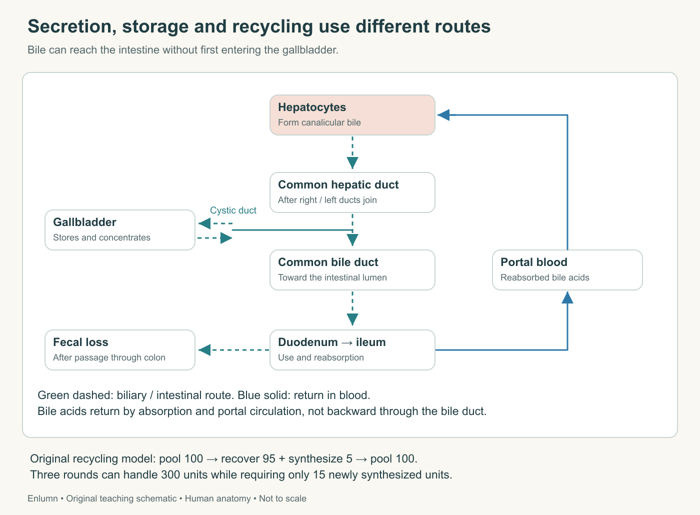
The cystic duct connects the gallbladder to the main biliary route. Bile can proceed to the intestine without storage. Reabsorbed bile acids return through portal blood, whereas unrecovered material continues through the large intestine before fecal loss. The recycling numbers are an original simplified model.
Recycling changes how we count production
Many bile acids return through enterohepatic circulation and are secreted again. The liver handles both newly synthesized material and recovered material. Consequently, the amount secreted into bile over a day is not the same as the amount newly synthesized that day. Counting every outward journey as a newly made molecule would greatly overstate synthesis in a recycling system.
In a fictional model, a pool of 100 units completes three rounds. Each round loses 5 units and receives 5 newly synthesized units before the next round. Total secretion over the three rounds is 300 units; new synthesis is 15. The pool can remain at 100 throughout the sequence despite a cumulative throughput three times its size. These are arbitrary teaching units, not a recommendation or estimate of human bile-acid turnover.
Now imagine recovery falls to 85 units per round while replacement remains 5. The next pool has 90 units, then declines further if the conditions persist. Increasing synthesis might partially compensate, but the maximum possible response and the health of the producing cells matter. A recycling failure and a synthesis failure can therefore affect the same downstream supply through different routes.
This distinction also clarifies an otherwise puzzling combination: bile acids can be useful substances and still need regulated elimination. Retaining every molecule indefinitely would not be the same as maintaining an appropriate pool. Production, reuse, transport and loss must be considered together. A product's usefulness depends on its amount, location and timing, not on an unconditional label of good or bad.
Check your understanding: Why does a fall in the measured concentration of a liver-produced protein not prove that the liver is making less of it?
Expected answer: Concentration reflects amount divided by fluid volume, and the amount reflects synthesis, distribution and removal. Increased loss, altered distribution or dilution can lower concentration despite unchanged synthesis. Establishing a production change requires evidence that distinguishes those processes.
Application
Allow 12–15 minutes. Trace one albumin molecule from synthesis to blood and a bile acid from synthesis through secretion, intestinal use and return. Mark the different cell surfaces and fluid compartments involved.
Complete the protein-pool and bile-recycling calculations. Then write 150–200 words explaining why bile salts and lipases cooperate, and why a micelle is not a chylomicron. Your account should distinguish physical dispersion, chemical breakdown, uptake and transport after uptake.