One organ, two kinds of output
A bowl of rice, beans and vegetables has reached the stomach. Some of its contents will soon enter the duodenum. The pancreas participates in what happens next through two quite different routes. One delivers substances into the intestinal lumen, where they help make the meal digestible. The other delivers signals into circulation, where they influence how tissues handle available nutrients. Confusing those routes makes almost everything else about the organ harder to understand.
Locate the pancreas in three dimensions
The pancreas lies deep in the upper abdomen, mostly behind the stomach. Its broad head sits within the curve of the duodenum, the first segment of the small intestine. A short neck leads into the body, which extends toward the person's left, and the narrower tail approaches the spleen. These are regions of a continuous organ, not separate glands with independently assigned jobs. Endocrine and exocrine tissue are distributed within it.
Most of the pancreas is retroperitoneal: it lies behind the lining of the main peritoneal cavity. The tail has a different relationship to the peritoneal folds near the spleen. “Behind the stomach” therefore establishes an important spatial relationship but does not mean the pancreas lies inside the stomach wall. Nor does a front-view diagram show how deeply one structure lies behind another. The OpenStax anatomical overview supplies the main regional landmarks; its two-dimensional outline needs this depth qualification.
Imagine lifting the stomach forward in a simplified anatomical model. The pancreas becomes easier to see, but the act of moving a covering organ has changed the view, not the normal location. This matters when comparing illustrations. An exposed pancreas and a pancreas shown faintly behind the stomach may depict the same arrangement. Before deciding that two drawings disagree, identify what each has removed or made transparent.
Major blood vessels also pass close to the pancreatic neck and head. The portal vein forms behind the neck from large venous tributaries, while other vessels supply the pancreatic tissue through branching routes. You do not need to memorize every branch to understand the functional distinction: incoming arterial blood nourishes the gland, its capillaries exchange with tissue, and venous blood carries released hormones onward toward the liver. The duct system is a separate network.
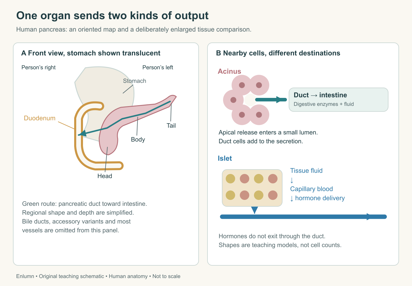
The oriented panel places the pancreatic head within the duodenal curve and the tail toward the person’s left. The stomach is shown translucent to expose the deeper organ. The enlarged tissue comparison separates a duct lumen from a vascular destination. Cell shapes and colors identify teaching components; they do not reproduce a histological cell count or the exact branching of a human specimen.
Follow the output that enters a duct
The exocrine pancreas releases a fluid containing digestive enzymes and ions into ducts. The smallest units include acini, clusters of secretory cells arranged around small lumens. Each acinar cell has a surface facing the lumen and other surfaces facing neighboring cells and the surrounding tissue. That orientation provides a direction for secretion: material is released toward the duct route rather than indiscriminately into every nearby space.
Tiny duct segments connect to larger ducts within and between tissue lobules. Much of the secretion ultimately enters the main pancreatic duct, which travels along the organ toward the head and opens into the duodenum. Commonly its terminal route joins the common bile duct near the intestinal wall. The resulting shared region and its muscular control are close neighbors, not synonyms: an ampulla is a widened passage, whereas a sphincter is muscle regulating an opening.
There is variation in how pancreatic ducts join and where they open. An accessory duct may provide another route into the duodenum. A useful first diagram can show the common arrangement while labeling it as such. It should not imply that every human has an identical branching pattern or that a second duct necessarily carries half the output. Anatomical presence, patency and functional contribution are separate questions.
Follow one secreted enzyme through this route. It begins as a protein made by a cell, is packaged and released into an acinar lumen, travels with fluid through ducts, and reaches the small intestine. It does not travel through the bloodstream to find rice in the stomach. Once in the intestinal lumen, its relevant substrate must actually meet it under suitable conditions. Production, delivery and effective action are three different achievements.
Follow the output that enters blood
The endocrine pancreas consists of clusters called pancreatic islets, or islets of Langerhans. These contain several hormone-producing cell types near an extensive capillary network. Beta cells release insulin; alpha cells release glucagon; delta cells release somatostatin. These signals have distinct effects and interact locally as well as participating in wider regulation. The islet is not simply a storage sack of insulin.
An endocrine cell releases its product into the surrounding extracellular space, from which the hormone enters capillary blood. The phrase “secretion into blood” is useful shorthand for this short sequence; the cell is not a pipe directly fused to a red blood cell. Capillary structure, tissue perfusion and the properties of the released molecule all matter to its movement. Hormones then reach tissues whose receptors and internal machinery permit a response.
The human endocrine pancreas atlas identifies islets as paler cellular regions within the more strongly stained exocrine tissue. Its description also makes an essential limitation explicit: routine hematoxylin-and-eosin staining does not reliably identify each individual endocrine hormone type. A pale islet is not a photograph of insulin molecules, and a dark nucleus is not sufficient evidence that a cell is a beta cell.
Insulin does not enter the pancreatic duct to digest carbohydrate. It is a peptide signal that changes activities in responding tissues. Likewise, a pancreatic digestive protease is not the signal telling skeletal muscle to increase glucose uptake. Both are proteins made by pancreatic cells, yet their destinations and functions differ. Chemical category alone does not determine physiological role; location and interacting partners complete the explanation.
Read a real tissue section
Open the atlas and choose Islet of Langerhans 1. The inspected view shows a broad, relatively pale region with many purple nuclear profiles and small intervening channels, surrounded by darker, clustered exocrine tissue. The boundary is irregular rather than a perfect circle. Some channels contain pink-red material consistent with blood-cell profiles. Use the atlas's annotation to locate the islet, then ask what the image itself makes visible.
You can describe relative staining, cellular organization and the presence of small spaces. You cannot watch a hormone being released in a fixed section. Nor can you infer the islet's full volume from the area of this slice. A section through the edge of a large three-dimensional object can look smaller than a central section through a modest one. The shape problem is geometrical, not a defect in microscopy.
Now open the exocrine view of the same human specimen and select Centroacinar Cells. The inspected field shows tightly arranged acinar profiles with dark nuclear regions and differences in staining toward the small central spaces. The atlas identifies pale duct-associated cells within the central acinus. Centroacinar names their position at the beginning of the duct system; it does not mean they are endocrine cells at the center of an islet.
The atlas also warns that red blood cells found in some ducts are a postmortem artifact. That warning prevents a plausible but incorrect inference: seeing a red cell in a preserved duct would not establish that blood normally shares the pancreatic secretion route. Preparation can alter appearances. A sound reading combines visible evidence, the annotation, knowledge of the method and awareness of artifacts, without allowing any one of those to substitute for all the others.
Cellular polarity creates a useful direction
An acinar cell's basal region contains abundant machinery for producing secreted proteins. Its apical region, facing the lumen, contains secretory granules. Polarity means that different cell surfaces and internal regions have different organizations. It is a maintained biological property, involving membrane sorting, junctions and transport, rather than merely a convenient label for the top and bottom of a drawing.
Picture a room with a preparation bench on one side and a serving hatch on the other. The analogy helps identify a directed process, but the cell has no cook deciding where to stand. Molecular signals, membrane proteins and cytoskeletal transport organize its work. If a diagram uses the same arrow for protein synthesis, movement of a granule and flow down a duct, add labels. Those arrows represent different physical events even though they form a connected sequence.
Duct cells also have polarity. Their membrane transport can add bicarbonate-rich fluid to the lumen, while other membrane surfaces exchange with the tissue environment. As Hart and Conwell's human secretion review explains, these cells contribute to the secretion itself. A pancreatic duct therefore cannot be understood entirely as passive plumbing. Losing a transport function can impair delivery and conditions for digestion even when the passage remains anatomically open.
Endocrine cells use regulated secretion too, but their relationship to nearby capillaries serves a different destination. The shared machinery of protein production and vesicle release does not erase the distinction. A useful drawing puts a duct lumen beside an acinus and a capillary beside an islet, with separate labels for extracellular space. It should never draw an islet as a bulb attached to the exocrine duct.
Why proportions do not tell us importance
Exocrine tissue occupies most of the pancreatic tissue visible in ordinary sections, while islets occupy a much smaller fraction. That observation does not mean the endocrine role is correspondingly unimportant. A small amount of a signal can influence a large responding tissue mass because the signal alters ongoing cellular processes. Hormone molecules are not providing the bulk material or energy needed for every response they coordinate.
Use an invented tissue map containing 960 area units of exocrine profiles and 40 of endocrine profiles. The endocrine area fraction is 40/1,000, or 4%. This is a measurement of the supplied map. It is not automatically the organ's volume fraction, the fraction of cells, the fraction of blood flow or the fraction of physiological importance. Converting one into another requires additional assumptions or measurements.
Even the cell count would require more information. A region occupied by relatively large cells can have a different number of cells than an equally sized region of smaller ones. Spaces, capillaries and connective tissue contribute area too. This is why we will use histology to explain arrangement without pretending a single attractive field provides a complete quantitative census of the pancreas.
Test the separation with a fictional obstruction
Suppose a supplied model has intact islets and normal blood supply, but the main pancreatic duct has become obstructed near its outlet. The immediate pathway problem concerns exocrine delivery. Enzymes and fluid made upstream cannot reach the intestine normally through that route. It does not follow that insulin has lost its exit, because insulin normally leaves by the vascular route. That is the first prediction of the model, not a complete forecast of a disease.
Over time, obstruction can affect tissue and therefore have consequences beyond the original route. We have deliberately held those later changes aside to isolate the first mechanism. Good simplification states what it has held constant, then recognizes when those assumptions would fail. “The endocrine route is separate” is a sound anatomical claim; “a duct problem can never affect endocrine function” is an unjustified extension.
Reverse the model. If beta-cell insulin secretion is impaired while acinar and duct function are initially preserved, digestive enzymes may still reach the meal. Glucose can be generated by digestion and absorbed into blood even though later regulation of its use is disrupted. This distinction will organize the entire course: the intestine prepares and takes up materials; endocrine signals help coordinate what happens after and during their arrival.
Check your understanding: Why would an isolated pancreatic duct obstruction not immediately prove that insulin cannot leave the pancreas?
Expected answer: Insulin is released from islet cells into tissue fluid and capillary blood, whereas exocrine secretion uses the ducts. The supplied obstruction directly impairs the duct route; broader endocrine effects would require additional changes to tissue or perfusion.
Application
Allow 15–20 minutes. Draw the pancreas beside the duodenum and mark the person's right and left. Add two enlarged insets: an acinus connected to a duct, and an islet associated with capillaries. Trace one digestive enzyme and one insulin molecule from their cells to their next destination.
Use the linked human atlas to write three observations and two limits of interpretation. Include the distinction between a two-dimensional section and a three-dimensional islet. Finish with 150 words explaining the fictional duct obstruction. Keep this drawing for the final course task; success depends on separate routes, correctly placed boundaries and an explicit distinction between immediate and later consequences.