Repair and Altered Structure
Two marks have the same outline on a diagram of a forearm. One removes only superficial epidermal material. The other extends through the dermis and destroys structures beneath the outline. Calling both “a small skin injury” preserves their surface size while discarding much of the information needed to understand their consequences. Area tells us how much boundary is involved; depth tells us which parts of the organ have been disrupted.
Repair brings the course's separate functions together. A disturbed barrier changes exchange with the environment. Injured vessels can leak. Sensory endings and immune signals alter communication. Surviving epithelial cells, connective tissue cells, and vascular cells contribute different pieces of reconstruction. The outcome depends on coordinating those contributions, not simply producing more cells as quickly as possible.
Start with a section, not a calendar
A superficial injury confined to the avascular epidermis need not bleed. If an injury reaches and disrupts dermal vessels, bleeding becomes possible. This does not mean that absence of visible blood measures exact depth: vessel involvement, local conditions, and observation all matter. It means that locating blood vessels prevents the mistaken idea that every layer contains the same structures.
In a partial-thickness injury, some dermis remains. Depending on depth and site, surviving epithelial portions of appendages can remain within the affected area. A full-thickness skin injury destroys the epidermis and full depth of dermis at that site; deeper tissue may also be involved. These descriptions identify anatomical extent, not a complete treatment decision. Rittié's review of repair mechanisms explains why surviving structures influence the routes available for resurfacing.
Consider an original model of a square injury measuring two centimeters on each side. Its surface area is four square centimeters whether it is shallow or deep. Give the shallow version a surviving network of epithelial structures beneath its surface and remove that network in the deep version. The same surface outline now encloses different starting conditions. A drawing that omits depth would conceal the difference before any repair process begins.
Even wounds with matching dimensions can differ in tissue viability, contamination, mechanical conditions, and blood supply. Geometry is necessary information, not sufficient information. The goal is to build a causal description in layers: what was lost, what survived, what must be restored, and what conditions support or interfere with restoration.
Stopping a leak and building a temporary platform
When vessels are injured, hemostasis limits blood loss. Platelets adhere and aggregate at the damaged site, while coagulation produces fibrin that helps stabilize the clot. A platelet plug and a fibrin network are related components, not interchangeable names for one material. Local vascular responses also contribute to limiting loss.
The clot has an immediate mechanical role, but the injury environment is already doing more than plugging a hole. Cells release signals that influence recruitment and activity of other cells. Temporary extracellular material provides a setting through which cells interact and move. This provisional arrangement is different from the mature architecture of intact dermis. The wound-healing physiology overview describes the overlapping processes of hemostasis, inflammation, proliferation, and remodeling.
A temporary bridge offers a bounded analogy. It may restore passage before the permanent supports are complete. Success at the first job does not prove completion of the second. Similarly, stopping a visible leak is not evidence that tensile strength, epidermal permeability, sensory function, and appendages have all returned to their earlier state.
The analogy also has a limit. Biological repair does not involve a separate engineering crew following an external blueprint. Cells respond to local signals and mechanical conditions while altering those same conditions. Their work changes the environment that guides subsequent work. Repair is a regulated interaction, not the assembly of inert replacement parts.
Inflammation has work to do
Inflammatory activity recruits and coordinates defenses and the handling of damaged material. Neutrophils and macrophages contribute through different, overlapping activities, including engulfment, signaling, and interactions with extracellular material. The population and behavior of cells change over time. A simple diagram showing one cell type disappearing at midnight when the next arrives would misrepresent that overlap.
Inflammation following injury is not identical to infection. Tissue damage can initiate inflammatory signaling without a microbial infection being the cause. Conversely, microbial presence at an exposed surface does not by itself establish tissue invasion. These distinctions continue the second chapter's separation of resident organisms, barrier breach, and immune response.
Imagine three fictional measurements collected from a repair site: the number of a selected immune cell, the concentration of a signaling molecule, and the amount of intact matrix. An increase in the first measurement cannot, by itself, tell us whether the overall response is useful, excessive, resolving, or ineffective. We need the cell's activity, the surrounding conditions, and the later outcome. A count is evidence about abundance, not a complete verdict on function.
This is why “more inflammation means better repair” fails as a general rule. A process can be necessary at one level while persistent or poorly controlled activity interferes with later reconstruction. Equally, “all inflammation must be absent before anything grows” is wrong. Repair processes overlap in both time and space. One part of a wound may be undergoing epithelial advance while another still contains substantial inflammatory activity.
Closing the surface and rebuilding underneath
Reepithelialization means restoring epithelial coverage. Keratinocytes change their behavior, migrate, proliferate, and eventually reestablish a stratified surface. Cells near wound margins can contribute; surviving epithelial appendages can provide additional sources in appropriate injuries. A continuous new covering still needs maturation and attachment. Coverage and fully recovered barrier performance are not synonymous.
Fibroblasts contribute extracellular matrix, while new vascular growth helps support active tissue. Granulation tissue is the vascular, cellular connective tissue that develops during repair; it is not the epidermal stratum granulosum despite the similar word. Epithelial coverage lies above a wound bed whose structure is also changing. Schultz and colleagues' account links these processes to subsequent matrix remodeling.
An original two-map exercise makes the distinction visible. On the first map, shade every point where epithelial coverage is present. On the second, mark where the underlying tissue has recovered its former organization. The maps need not match. One could show a continuous surface while the other still shows altered collagen arrangement, vessels, or appendage loss. “Closed” answers one important question; it does not answer every question about recovery.
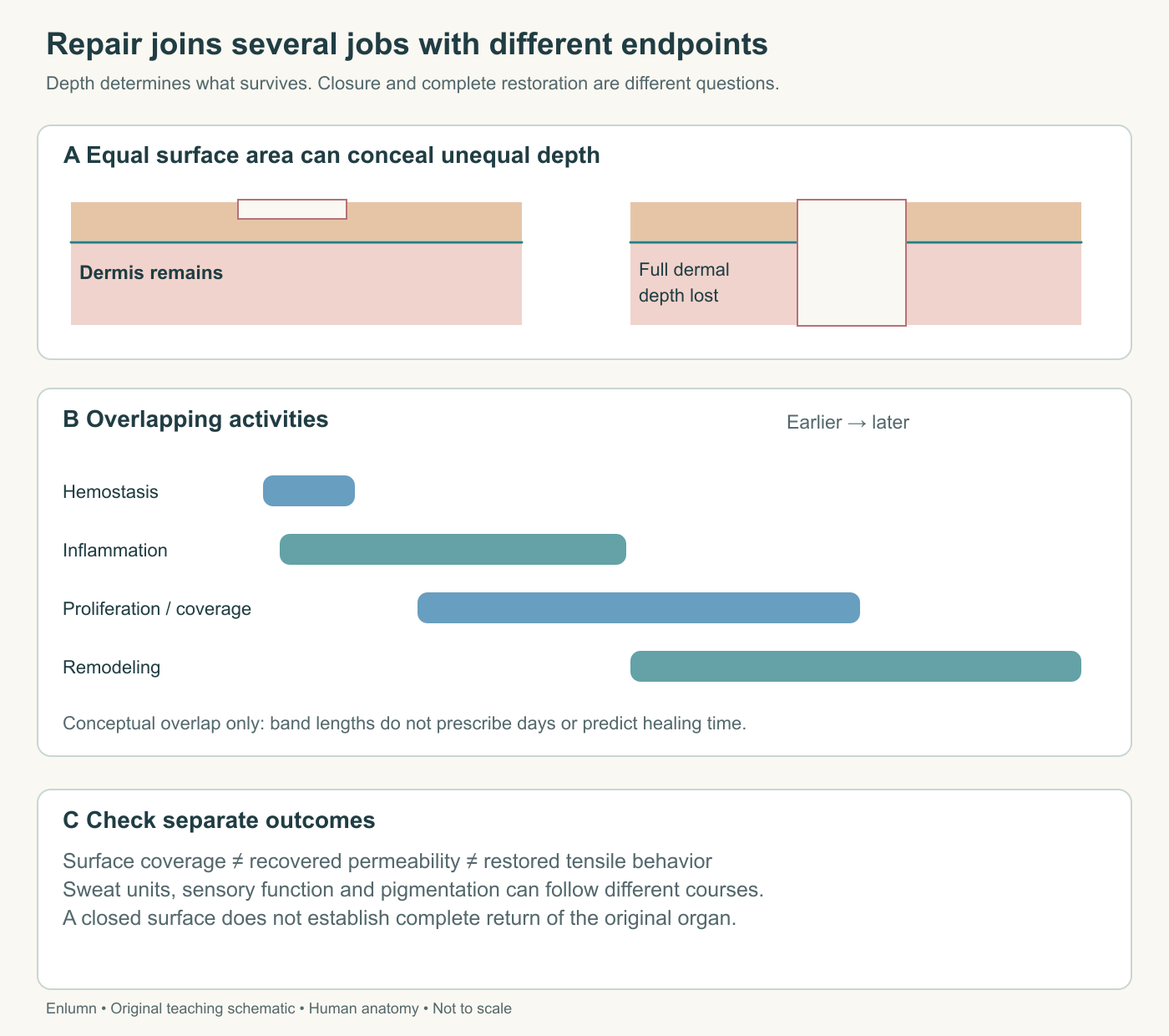
The diagram's time bands have no numbered days. Their purpose is to show overlap and different endpoints. It would be a mistake to convert the drawing into a timetable for judging an individual wound. Location, extent, health, and the circumstances of injury can change the course of repair.
What a human study adds
A study by Rittié and colleagues examined repair after controlled partial-thickness injury in healthy human forearm skin. The researchers used tissue samples, immunohistochemistry, and three-dimensional reconstruction to investigate the sources of new epithelial outgrowths. They found contributions associated with eccrine sweat structures as well as hair-associated units. The result adds a repair role to the sweat apparatus studied earlier for secretion and heat exchange.
This evidence concerns a particular experimental injury and surviving structures. It does not show that a completely destroyed gland regenerates intact, that sweating itself closes wounds, or that every body site repairs identically. The techniques and research injuries are not activities to reproduce. The useful conclusion is anatomical: structures extending below the surface can help supply cells for restoring that surface when appropriate portions survive.
Now use an explicitly fictional geometry model. A one-dimensional gap is ten millimeters wide, with growth sources only at the two ends. Each advancing front would have to cover five millimeters before meeting at the center. Add surviving sources at two-millimeter intervals. In the simplified model, neighboring fronts now need to cover only one millimeter each.
This calculation predicts a shorter distance, not an exact healing time. It assumes viable sources, a suitable substrate, and symmetric growth. Cell behavior, wound-bed conditions, and three-dimensional structure remain unmodeled. Its value is to reveal why distributed starting points can matter independently of total surface area. The study supplies evidence for an anatomical contribution; the invented line model isolates one possible geometrical consequence.
A further inference would require further evidence. For example, showing that an outgrowth lies near a gland is different from identifying the precise lineage of every contributing cell. Three-dimensional reconstruction improves spatial interpretation, but a research method answers the questions its design can support. Reading a study well includes resisting attractive claims that go beyond its observations.
Matrix quantity is not matrix organization
Collagen and other extracellular components are continually produced, modified, and removed during remodeling. Changes in organization and cross-linking influence mechanical properties. Tensile strength describes resistance to being pulled apart; it is not the same as flexibility, thickness, or impermeability. A repaired region can improve in one property while differing from its original condition in another.
Consider two fictional strips, each containing the same mass of reinforcing fibers. In strip A, fibers bridge the direction of the applied pull. In strip B, many lie in orientations that contribute less to resisting that pull. Equal mass does not guarantee equal load-bearing behavior. Real skin has a complex network responding to multiple directions, but the model explains why counting collagen alone cannot establish restored mechanics.
Now distinguish force from stress. A model strip carries ten newtons over a cross-sectional area of two square millimeters, giving five newtons per square millimeter. Another carries the same force over four square millimeters, giving 2.5. The same applied force produces different stress because the area differs. A claim that one specimen “held ten newtons” needs dimensions and testing conditions before it becomes a fair material comparison.
These numbers are supplied engineering examples, not measurements of wounds. They show why statements about strength require a reference: compared with which tissue, at which site, under which loading direction, and at what stage? A single universal percentage of “normal strength” would hide those conditions. Looking closed is even farther from being a direct mechanical measurement.
Some repair-associated cells develop contractile behavior and can help draw tissue inward. Contraction can reduce the area requiring coverage; excessive contraction can limit movement. This connects a local skin change to the mechanics of a joint or opening. The word “repair” therefore does not guarantee restoration of the exact shape or movement that existed before injury.
A scar records reconstruction with differences
A scar is a structural outcome of repair, especially when dermal tissue has been disrupted. It is not simply dried blood left on the surface. Its collagen organization, pigmentation, sensory properties, and appendages can differ from the original tissue. A superficial disturbance that restores epidermis does not inevitably produce a permanent dermal scar.
The American Academy of Dermatology's keloid overview distinguishes a keloid, which grows beyond the original wound boundary, from a hypertrophic scar, which remains within that boundary. This is a useful contrast in growth pattern, not a photograph-based diagnostic exercise. Color alone cannot establish either category, and population patterns do not determine what will happen to one person.
Suppose a fictional repaired region has a continuous surface and little water loss under the specified test conditions, but no functioning sweat gland in its center. Barrier recovery and local secretory recovery have separated. In another case, coverage is present but the region stretches differently from nearby skin. Again, one endpoint has improved without every feature returning.
These examples do not imply that a small local alteration necessarily changes whole-body temperature regulation or overall movement. Scale matters. Losing one tiny patch of function differs from losing a broad area. The correct explanation links local structure to a local consequence first, then asks whether the affected extent is large enough to produce a wider effect.
Constructing a complete explanation
Return to the two equally sized marks. For the shallow version, describe disruption of the surface barrier and the surviving cells available for renewal. Do not invent vessel injury when the supplied section shows none. For the deep version, add the lost dermal matrix, affected vessels, and destroyed appendages actually shown. Then explain which repair tasks each loss creates.
A complete account should follow at least three chains. One connects barrier disruption to altered exchange and the need for renewed epithelial organization. Another connects tissue injury to local vascular and immune responses. A third connects matrix and appendage loss to reconstruction and possible lasting functional differences. These chains interact, but they are not interchangeable.
The five chapters have treated skin as an organ whose boundary is actively maintained. Layered structure supports exchange control; nerves and vessels connect it to wider regulation; pigment changes how radiation interacts with tissue; repair reconstructs functions after disturbance. The final test is whether you can use those relationships to explain a new section drawing without falling back on “skin protects us” as the whole answer.
Check your understanding: A fictional wound has regained continuous epithelial coverage. Does that establish restored tensile strength, normal sweating, and identical pigmentation? Explain using tissue location and different repair endpoints.
Expected answer: No. Coverage describes the surface. Dermal matrix organization and mechanics may still be changing, destroyed sweat structures may remain absent, and pigment handling can follow a different time course. Each function needs relevant evidence rather than inference from closure alone.
Application
Allow thirty to forty minutes for the course capstone. Write 600–900 words accompanied by an annotated section. Use this supplied case only: an injury interrupts the epidermis, damages part of the dermis and a small vessel, leaves some epithelial appendages alive, and destroys one complete local sweat unit. Later, epithelial coverage returns but the connective tissue has a different organization. Do not examine, photograph, or experimentally injure skin.
Your section should identify epidermis, basement-membrane region, dermis, the vessel, surviving epithelial structures, the lost sweat unit, and underlying tissue. Explain three immediate consequences and the repair work associated with each. Then distinguish what surface closure establishes from what it leaves unresolved about exchange, sensation, mechanical behavior, pigmentation, and local sweating.
Include one calculation from the chapter's supplied geometry or stress models, state its assumptions, and explain why it cannot predict an individual's healing time. Use the human repair study to support one bounded anatomical claim. End with two observations that would discriminate between competing explanations of a fictional outcome.
A strong capstone links every claim to a structure or measurement, shows overlapping processes rather than rigid day gates, distinguishes repair from complete restoration, respects the limited size of the affected area, and avoids diagnosis or treatment recommendations.