Where absorbed materials go
The glucose unit from the pasta and the long-chain fatty acid from the olive oil have reached an enterocyte, but their stories now diverge. Glucose can leave toward local blood capillaries. Much of the absorbed long-chain lipid is rebuilt and packaged before entering intestinal lymph. Both pathways eventually connect with circulating blood, yet their first destinations, molecular forms and intervening steps differ.
This chapter follows those differences beyond the intestinal wall. The objective is not to memorize every named vessel. It is to produce a route that preserves three facts: the epithelial cell is an active processing compartment; blood and lymph are distinct local pathways; and entering systemic circulation is not the same as having already passed through the liver's portal supply.
Portal blood makes the liver an early destination
After crossing an enterocyte, a glucose molecule enters tissue fluid and can cross into a nearby capillary. The capillaries drain into venules and larger intestinal veins. Much of this drainage reaches the superior mesenteric vein and then the hepatic portal vein, carrying blood to the liver. The portal arrangement connects intestinal capillary beds with another specialized vascular bed in the liver before blood returns to the heart.
The word portal describes that organization, not a special kind of nutrient molecule. The same blood carries water, ions and many other substances, and it includes blood that entered the intestine through its arterial supply. Nutrients added from the lumen become part of a preexisting circulation. The intestine does not manufacture a new volume of blood whenever a meal arrives.
In the liver, blood enters sinusoids, where substances can exchange with hepatic cells. It then leaves through hepatic veins toward the inferior vena cava and the right side of the heart. The Liver course examines storage, conversion and release in detail. Here, the essential implication is that the liver can modify the material reaching the rest of the circulation after intestinal absorption.
Not every glucose molecule is necessarily retained on its first passage, and passing through a vascular bed does not mean entering every cell within it. Some material proceeds onward; some is taken up or transformed. A route map describes opportunities and order. It does not, by itself, specify the fraction processed at each stop.
Net appearance is a balance
Imagine an isolated fictional intestinal region with blood flow of 10 volume units per minute. The entering arterial concentration of a selected substance is 4 amount units per volume, and the leaving venous concentration is 7. Assuming equal inflow and outflow volumes, net addition to the blood is 10 × (7 − 4), or 30 amount units per minute.
Multiplying 10 by the venous concentration alone gives 70 units per minute, but 40 were already arriving in arterial blood. The difference matters. The organ's contribution is not identical to everything leaving it. In real research, investigators also account for the substance measured, changing conditions and the limits of their sampling and flow estimates.
Now suppose that 40 labeled nutrient units cross into the intestinal wall during the minute, but the tissue retains or transforms 10 before venous departure. Net venous appearance of the measured form is 30. The blood measurement underestimates entry across the epithelium if it is interpreted as a direct measure of that earlier event. Enterocytes use nutrients themselves and can transform them, so the wall cannot always be treated as an inert connector.
Reverse the situation: tissue releases some material previously stored or made within it while current luminal uptake is small. Venous appearance may then overstate the contribution of the current meal. The proper question is always specific: appearance of which substance, from which source, across which boundary, during which interval?
A long-chain lipid is rebuilt
The fatty acid from olive oil reached the apical membrane after luminal digestion and exchange from the micellar environment. Uptake can involve both membrane partitioning and protein-mediated processes. It is misleading to say that every lipid simply passes anywhere through a membrane without cellular participation. Inside the enterocyte, much of the absorbed long-chain fatty-acid material is used to re-form triglycerides.
Re-esterification links fatty acids into ester-containing molecules again. Why break a triglyceride down only to build triglycerides afterward? Because the requirements for crossing the luminal boundary differ from the requirements for transporting large amounts of lipid in an aqueous circulation. Digestion creates accessible components; intracellular assembly creates a suitable transport form. The final molecule need not have the exact same partners as the original dietary triglyceride.
An original label exercise makes that last point clear. Imagine three triglycerides, each carrying one distinguishable fatty-acid label among their chains. After digestion, their components mix with other absorbed and cellular material. The three labeled fatty acids may enter three different newly formed triglycerides, or some may be used or retained by the cell. Recovering the labels does not prove recovery of the original three intact molecules.
Within the enterocyte, triglycerides associate with other lipids and proteins in lipoprotein particles called chylomicrons. Their assembly begins in the endoplasmic reticulum and involves subsequent processing and secretory transport. Apolipoprotein B48 is a structural component; microsomal triglyceride transfer protein contributes to the assembly process. These names identify machinery, but the central concept is packaging before export. The intestinal assembly section of Endotext describes these stages.
A micelle and a chylomicron are different objects
A mixed micelle in the intestinal lumen helps carry poorly water-soluble digestion products through a watery environment. A chylomicron is assembled within an enterocyte and exported across its basolateral surface. The first assists access to uptake; the second supports transport after uptake. Drawing one circle that passes unchanged from lumen into lymph erases both digestion and intracellular assembly.
Chylomicrons contain a hydrophobic lipid-rich interior and a surface suited to interaction with the surrounding aqueous fluid. They are dispersed particles, not triglycerides chemically converted into freely water-soluble molecules. A container can travel in water without its contents having the same solubility as the container's exposed surface.
Think of a fictional enterocyte receiving 90 units of long-chain fatty-acid material during an interval. Suppose 60 units leave in secreted particles, 20 remain in cellular lipid stores and 10 are used or transformed by the cell. All 90 crossed the apical boundary, but only 60 have reached the selected export route by the end of the interval. The numerical balance is invented to show why uptake and immediate secretion can differ.
If the stored 20 units are released later, a subsequent rise in lipid output does not prove new absorption at that later moment. Storage introduces a time delay between input and measured output. This distinction is useful whenever a post-meal blood curve is described as though it directly photographs what the intestine is doing right now.
Lymph supplies the first major route away
After basolateral release, chylomicrons pass through the extracellular space toward lacteals, the lymphatic capillaries within villi. The local lymphatic route accommodates these particles in a way the ordinary intestinal blood-capillary pathway does not. The two vessels are neighbors within the same supporting tissue, but they are not interchangeable entrances.
Intestinal lymph then moves through collecting vessels and lymph nodes toward larger lymphatic channels. Much of it reaches the thoracic duct, which normally joins the venous circulation near the junction of the left internal jugular and left subclavian veins. Anatomy varies, and a simplified diagram does not need every variant to preserve the usual route. The drainage should not be placed at the right subclavian vein as the standard intestinal pathway.
Lymph movement depends on pressure differences, vessel contractions, valves and surrounding movements. It is not pumped around a closed circuit by a separate lymph heart. Valves help constrain backward movement while forces generated in and around the vessels support forward flow. These mechanics explain why a drainage pathway is part of absorption's supporting system, not an optional final detail.
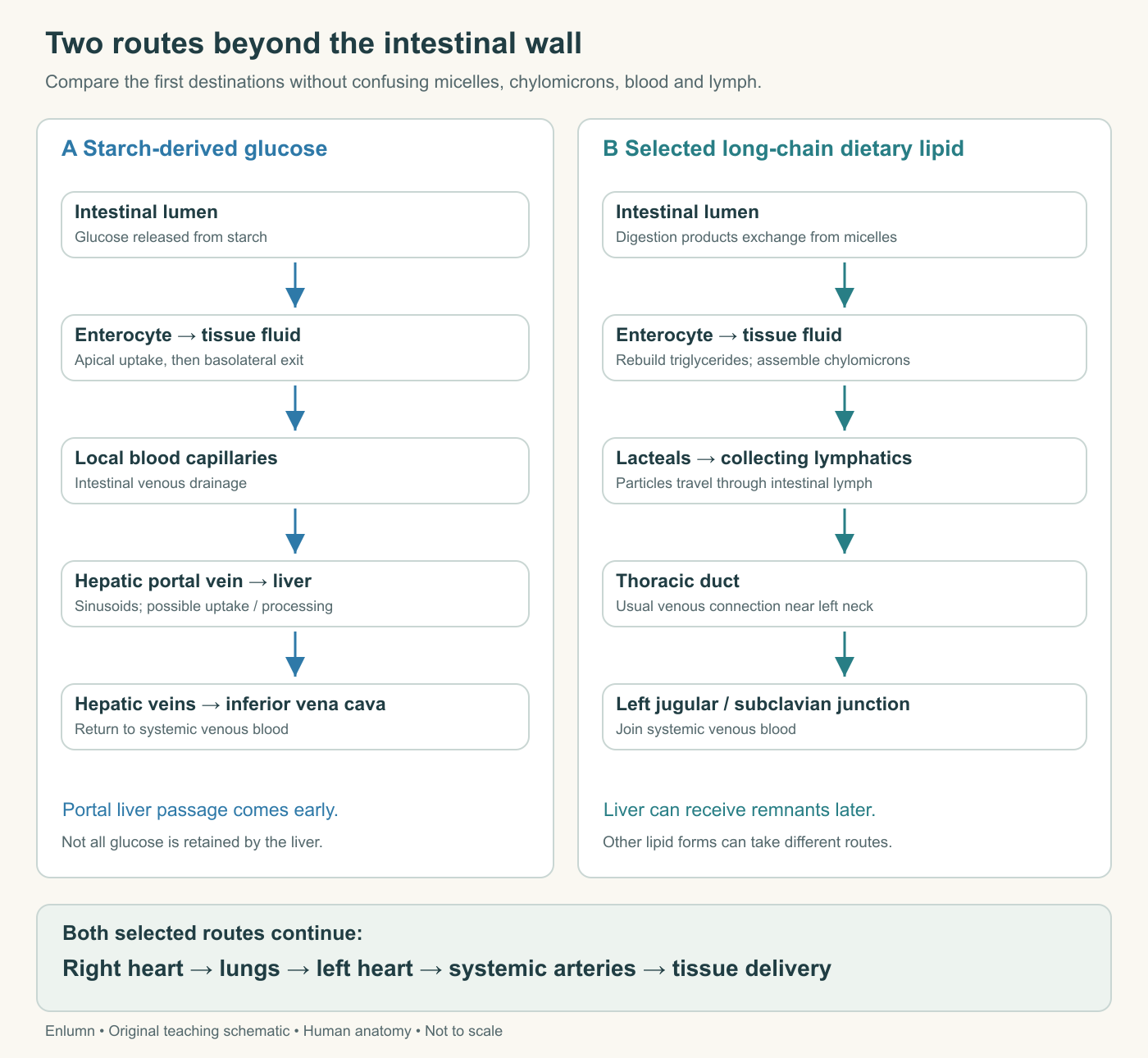
The glucose route reaches the liver through portal blood before returning to the heart. The selected chylomicron route reaches systemic venous blood through lymph. Both then pass through the right heart and lungs before systemic arterial delivery. Molecular processing, collateral vessels and anatomical variants are simplified.
The lipid route still connects with the liver later
Once lymph joins systemic venous blood, its particles travel toward the right heart, through pulmonary circulation, into the left heart and onward through systemic arteries. In suitable tissue capillaries, lipoprotein lipase acts on triglycerides carried by chylomicrons, releasing fatty acids that tissues can take up. This enzyme acts in a different place from pancreatic lipase in the intestinal lumen, despite the shared word lipase.
The particles change as they deliver lipid. Chylomicron remnants can subsequently be taken up by the liver. Thus, saying that this selected lipid route initially bypasses portal delivery is not saying that its material never reaches the liver. The difference concerns the order of early transport and processing.
Nor do all lipids follow the same route in the same proportion. More water-soluble short-chain and many medium-chain fatty acids can reach portal blood more directly. Chain length, molecular context and conditions matter. Our long-chain olive-oil example was chosen to make the chylomicron pathway clear, not to establish an exceptionless rule for every molecule described as fat.
Similarly, a drawing of glucose's portal route does not describe every detail of amino-acid, vitamin or mineral handling. Some compounds bind to carrier proteins; some are modified or retained by intestinal cells. A useful introductory comparison selects representative materials while naming the limits of the selection.
Recycling is another kind of journey
Bile acids offer a third route pattern. They are delivered into the intestinal lumen as digestive helpers and many are recovered, particularly through specialized uptake in the ileum, then returned toward the liver in portal blood. The liver can secrete them again. This enterohepatic circulation reduces the amount that must be newly synthesized to support repeated use.
Do not confuse the recycled helper with the dietary lipid cargo. They may share a micellar environment in the lumen but part company during absorption and subsequent transport. Following the helper explains conservation and replenishment; following the fatty acid explains nutrient uptake and distribution. One arrow cannot answer both questions unless it labels the material at every stage.
Use an invented recovery example. A pool contains 100 marked helper units, and each modeled cycle recovers 90 percent of those still present, with no replacement. After one cycle, 90 remain; after two, 81; after three, 72.9. Repeated reuse does not eliminate loss. The human body normally replenishes losses and regulates the system, so this declining sequence is a deliberately incomplete model.
Suppose instead that 10 units are added after each cycle. Starting from 100, losing 10 and adding 10 returns the pool to 100. That stable pool can coexist with substantial ongoing secretion, recovery and synthesis. A constant measured amount does not establish that the material is motionless or that no energy is spent maintaining it.
Compare two explanations of one blood curve
Imagine a graph showing a later-than-expected appearance of labeled dietary lipid in systemic blood. One explanation is delayed gastric delivery. Another is slower intestinal processing or export. A third is altered removal from blood, which affects the curve after entry. The graph alone does not distinguish these possibilities, because circulating concentration reflects both appearance and disappearance.
A better study might measure several compartments or combine a tracer with information about gastric emptying, particle production or clearance. The added measurements need to correspond to competing explanations. More data are useful when they locate the uncertain step, not merely when they repeat the same endpoint more frequently.
The same reasoning applies to glucose. A smaller systemic rise after a meal need not mean less glucose crossed the intestine. Hepatic handling, tissue uptake, endogenous glucose production and timing can all contribute. This course does not ask you to infer intestinal function from a personal blood reading. It asks you to recognize why that inference requires more than one number.
You can now follow the two meal components beyond the villus. The glucose route and the long-chain lipid route involve different molecular forms and early destinations, yet both depend on a maintained epithelial boundary, viable cells and onward flow. The final chapter examines what happens when the surface, its movement or its defense is altered.
Check your understanding: Why is “fat goes straight from the gut into the liver” an inadequate account of the selected olive-oil pathway?
Expected answer: Long-chain lipid products are taken up and largely rebuilt into triglycerides within enterocytes, packaged into chylomicrons, exported into intestinal lymph and carried through the thoracic duct into systemic venous blood. The liver can receive remnants later; other lipid forms may have different routes.
Application
Spend 15–20 minutes drawing glucose and a long-chain dietary fatty acid from lumen to systemic arterial blood. Label each change in molecular form and each named vascular or lymphatic destination. Distinguish the two lipases by location.
Then calculate net addition to blood when flow is 8 volume units per minute, arterial concentration is 3 and venous concentration is 7. State one reason this need not equal apical uptake.
Model interpretation: Net addition is 32 amount units per minute: 8 × (7 − 3). Intestinal metabolism, storage or release can separate venous appearance from entry across the apical membrane. The lipid drawing should show cellular assembly before lymph, and the glucose drawing should show portal passage through the liver before return to the heart.