Storing, mixing, and emptying
Imagine watching the stomach through a transparent window. A region of its wall narrows; contents move toward the exit; much of that material then moves back into the chamber. Has the stomach failed to transport the meal? Not necessarily. Backward movement within the stomach can contribute to processing while only part of the contents enters the intestine. To understand what you are seeing, you need three separate descriptions: how the chamber receives a meal, how it rearranges that meal, and how material actually leaves.
We will use a sequence of original models rather than a single universal digestion time. Real meals differ in composition and physical structure, and the stomach responds to information from the intestine. A bowl of liquid and a mixed solid meal are not interchangeable test objects. Even for one meal, emptying is a process unfolding over time, not an appointment at which the entire contents leave together.
Receiving more without squeezing harder
Accommodation is the adjustment that allows the stomach to receive a meal with a limited rise in internal pressure. It involves a reduction in muscle tone and an increase in compliance, especially in the proximal stomach. Tone is sustained muscle activity; compliance describes how much volume changes for a given pressure change under specified conditions. The gastric sensorimotor review distinguishes accommodation from other motor functions and their measurements.
In a simplified pressure–volume experiment, suppose chamber A accepts an additional one hundred volume units while its pressure rises by two units. Chamber B accepts the same volume increase with a pressure rise of ten units. Over those particular intervals, A has greater compliance: fifty volume units per pressure unit compared with ten. These are invented units, not human reference values. A pressure–volume curve need not remain linear as filling continues, so the comparison applies to the specified intervals.
Now ask whether A must have a thinner wall than B. The measurements alone do not establish that. An actively controlled muscular wall can change its tone, and its geometry also matters. The response of a living chamber is not determined solely by the stiffness of an inert material sample. This is why the familiar balloon comparison helps with expansion but becomes misleading if it implies that gastric accommodation is entirely passive stretching.
Neural pathways connect swallowing, gastric signals, and responses to intestinal contents with gastric relaxation and other motor adjustments. Vagal connections and enteric motor neurons participate; inhibitory signaling can relax smooth muscle. The gastrointestinal nervous-control reference describes this coordination. “The nervous system stimulates digestion” is too broad a statement to explain it: an effective response can require relaxation in one region and contraction in another.
Draw two adjoining regions of a model chamber. If both contract strongly at once, the incoming meal encounters a different mechanical situation from one in which the receiving region relaxes while a downstream region processes existing contents. The timing and distribution of activity matter. A single average measure of muscular activity across the whole organ would conceal this coordination, just as a city's average traffic speed can conceal a blocked intersection.
The organ changes shape in three dimensions
A human MRI study reconstructed stomach shape in sixteen healthy volunteers receiving a liquid nutrient meal. The investigators divided the reconstructed organ into seven regions and tracked changes in volume and landmarks. The abstract reports differing regional contributions during early and later filling and emptying, together with changes in position and shape. This is evidence that the stomach's response cannot be represented completely by scaling up one fixed outline. The study's abstract is the level of access used here.
Its design suggests questions worth asking of any imaging result. How were regional boundaries chosen? How was the organ's shape reconstructed from the images? Was the same region followed consistently as the stomach moved? Those questions do not invalidate the findings. They identify what must work for the measurements to answer the anatomical question. A reported regional volume is partly dependent on a reproducible method of defining that region.
The participants and meal also limit the inference. Sixteen healthy volunteers consuming liquid nutrients do not represent every age, disease state, posture, or solid meal. We can learn that regional deformation occurs without treating the reported pattern as a compulsory script for every dinner. The full paper would be needed to evaluate details beyond the abstract, and we have not reproduced its images or numerical results.
Here is a useful drawing exercise. Sketch two different shapes that preserve the same entry and exit. Divide both using the same anatomical landmarks. Could one region gain volume while another loses it even though total volume remains unchanged? Yes. Internal redistribution can do that. An increase in local volume is therefore not automatically evidence of additional swallowing, and a decrease is not automatically evidence that material crossed the pylorus. Total and regional quantities answer different questions.
A narrowing wave can create a backward jet
The distal stomach, including the antrum, contributes strongly to mixing and the reduction of food-particle size. A propagating contraction can move contents toward the pylorus. As the outlet narrows, some contents are driven backward within the stomach, producing retropulsion. Repeated deformation and fluid movement help mix gastric secretions with food and work on particles. Material passing into the duodenum is only part of the moving material.
To see why this is mechanically possible, imagine a flexible tube with a narrowed exit. Press a moving constriction along the tube toward that exit. Contents ahead of the constriction cannot all pass through the narrow opening at once. Pressure and available pathways redirect some flow backward around the moving material. This model omits the stomach's three-dimensional geometry and active control, but it demonstrates why “the wall moved forward” does not mean “every particle moved forward.”
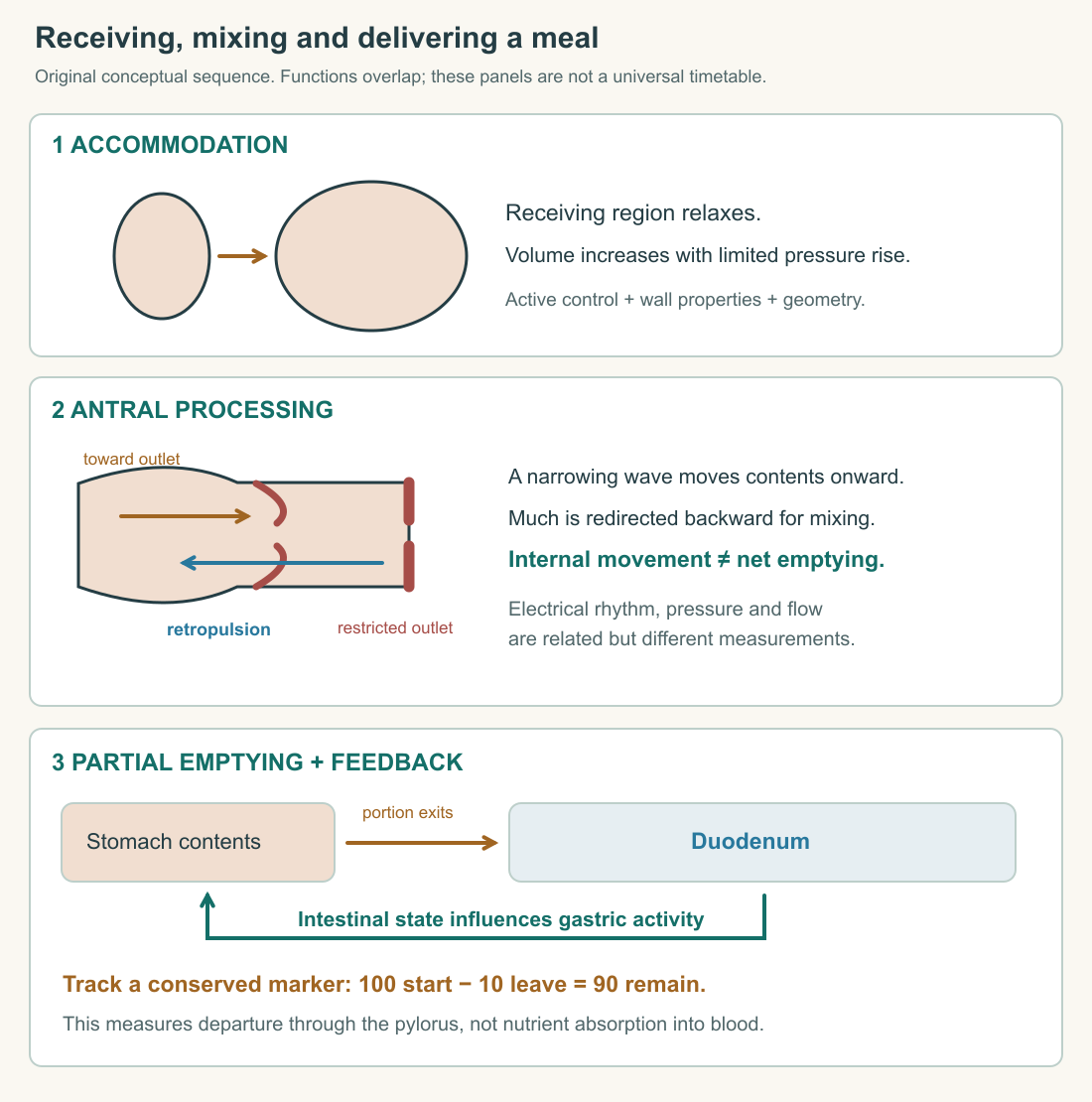
A simple marker ledger separates mixing from emptying. Put one hundred indestructible marker units in an imaginary chamber. During an interval, vigorous circulation moves eighty units from the proximal region into the distal region and sixty back again. Ten units cross the outlet. Ninety remain in the whole chamber. The gross internal movements can be much larger than net outflow. The ledger counts marker units, not real food particles that may fragment or dissolve.
If you instead tracked the number of visible particles, mechanical breakdown could increase that number even while food left the stomach. Four large pieces can become forty smaller pieces. A rising particle count would not demonstrate a growing amount of food. Choosing a conserved marker prevents that particular confusion. Real experimental markers also need validation: a liquid marker may not follow a solid meal identically, and attachment to food must be understood before interpreting the signal.
Electrical timing is not a contraction measurement
Interstitial cells of Cajal participate in generating and propagating gastric electrical slow waves. These rhythmic electrical changes organize the timing of muscle activity, but an electrical wave is not itself a measured displacement of food. The relation between electrical activity, muscle contraction, pressure, and transport includes several steps. The sensorimotor literature distinguishes electrical recordings, pressure measurements, accommodation measurements, and emptying tests because they observe different aspects of function.
Think of a row of lamps showing when motors receive a timing signal. Regular lamp flashes do not prove that every motor produces the expected force, that its linkage is intact, or that the load moves. Conversely, a load's final position cannot reconstruct every signal that occurred. The stomach is not a row of electric motors; the analogy identifies a problem in interpreting indirect measurements.
Suppose two model recordings show the same rhythm. One chamber develops strong coordinated pressure changes, while the other develops little effective deformation. The matching rhythm alone would miss the difference. Now suppose two chambers produce similar pressure peaks but have different outlet resistance. Their net outflow can differ. To explain a measurement, name its physical quantity and the additional relationships needed to connect it with your conclusion.
This is also why a quiet-looking surface image cannot settle a motility question. It may capture the interval between contractions, miss another region, or lack sufficient time resolution. The appropriate evidence is a sequence or a measurement designed to capture changing function. Anatomy provides the structure in which movement happens; physiology requires time as well as space.
The intestine helps set the delivery rate
The stomach's outlet is part of a coordinated junction with the duodenum. The intestine receives acidic, nutrient-containing chyme while pancreatic and other secretions contribute to subsequent processing. Its chemical and mechanical state feeds back on gastric activity. Fat and acid arriving downstream can slow further gastric delivery through neural and hormonal mechanisms. The gastric sensory and motor-function review describes the coupling between meal characteristics and emptying.
A useful comparison is a workshop feeding material to a second workshop. Sending material faster is beneficial only if the receiving stage can process the increased delivery. An upstream reservoir allows intake to occur in bursts while downstream work proceeds on a different schedule. The stomach's controlled delivery therefore serves the whole digestive sequence. Maximum outflow at every moment is not the objective.
Consider a stipulated downstream capacity of twelve processing units per interval. An upstream chamber releasing twenty units each interval creates a growing unprocessed load if all else stays fixed. Releasing ten units leaves capacity available. Those invented numbers illustrate a coordination problem; the intestine has no single fixed capacity corresponding to them. Its responses vary with substrate, secretions, absorption, and ongoing control. The model helps us ask what the feedback is coordinating without pretending to calculate a person's optimal emptying rate.
Solids and liquids also present different physical tasks. A digestible solid meal must be reduced and mixed before much of its solid component passes onward; liquid components can follow a different pattern. Consequently, an “emptying time” is incomplete unless we know the meal, the component tracked, and the endpoint. Time to the first detectable departure, time until half a marker has left, and time until very little remains are three different measures.
Read a curve without inventing a diagnosis
Here is an original marker example. At four equally spaced observations after the starting measurement, the amount remaining is eighty, sixty, forty, and twenty units from an initial hundred. The average loss over each interval is twenty units. We have stipulated no marker entry, production, destruction, or alternative exit. Under those assumptions the declining inventory measures cumulative departure through the outlet.
Now change the observations to ninety-five, eighty, forty-five, and twenty. The final amount is the same, but the interval losses are five, fifteen, thirty-five, and twenty-five units. One final image would conceal the different history. Half the marker leaves between the second and third observations, but the exact time cannot be known from these sparse observations without an interpolation assumption. Drawing a smooth curve through the points creates a model, not additional measurements.
Neither example supplies a clinical normal range. To compare with a reference population, we would need a defined meal, validated marker, sampling protocol, appropriate analysis, and knowledge of factors that influence the result. NIDDK's explanation of gastric-emptying assessment describes medically supervised measurements. The arithmetic here teaches the distinction between remaining amount and departure rate; it is not a home diagnostic test.
One more counterexample connects this chapter to the previous one. Suppose a marker-bearing liquid leaves normally but acid secretion differs. The outflow record does not establish identical chemical processing. Suppose acidity is similar but the solid component remains longer. The chemical measurement does not establish identical delivery. A complete account of the meal needs both chemical and mechanical descriptions, with their measurements kept distinct before their relationships are explained.
The stomach can now be understood as a receiving chamber, an active processor, and a regulated source of material for the intestine. Those descriptions overlap physically, but each gives us a different question to ask when function changes. In the last chapter we will use that separation to distinguish a damaged protective surface from impaired delivery, and to explain why a shared symptom does not identify a single mechanism.
Changing the observation interval changes what you can resolve. If two brief bursts of outflow occur between widely spaced images, the images may show only their combined effect. More frequent observation can reveal timing, but a more detailed record still needs a valid marker and a clear anatomical boundary. Precision in the time axis cannot repair a marker that follows the wrong component of the meal. Before improving an instrument, decide which question it is supposed to answer. “How often does the wall deform?”, “How much liquid crosses the outlet?”, and “How much of the solid meal remains?” require related but distinct observations. The best measurement is the one whose physical meaning matches the claim.
Application
Draw three snapshots of a meal: accommodation, antral mixing with retropulsion, and partial emptying. Use one arrow style for content movement and another for intestinal feedback. Plot the two invented marker series on the same axes and explain what a single final observation would miss.
Check your understanding: A chamber has large back-and-forth internal flows but loses only ten of its initial hundred conserved marker units through the outlet. How many remain, and does their retention imply that no useful movement occurred?
Expected answer: Ninety remain under the stated assumptions. Large internal movements can contribute to mixing and processing while net outflow stays small; movement within a chamber and departure from it are different quantities.