Getting oxygen across a surface
A fish opens its mouth, moves the region behind its head, and opens its mouth again. It is tempting to call each visible cycle a breath and assume that counting the cycles tells us how much oxygen it obtains. But the count leaves several questions unanswered. How much water moved? Where did it go? How much oxygen crossed from that water into blood? And how much of the oxygen reaching the blood eventually supported the working tissues?
A gill is an exchange surface supplied by two moving fluids. Water delivers dissolved oxygen to one side; blood carries oxygen away from the other. The fluids remain separated by living tissue. Understanding this arrangement means keeping ventilation, diffusion, circulation, and tissue use distinct while explaining how they cooperate.
Look inside the exchange region
In a generalized teleost, the large ray-finned group containing salmon and garibaldi, gill arches support filaments bearing thin exchange structures called lamellae. Water passes through spaces beside these surfaces while blood flows within them. The arrangement provides extensive contact across a short tissue barrier. A gill cover, the operculum, overlies the region, but the cover is not itself the principal oxygen-exchange surface. OpenStax: fish gill surfaces
Draw three different scales rather than one impossible close-up. At body scale, locate the mouth and the outlets behind the gill region. At organ scale, show the supporting arch and repeated filaments. At exchange scale, separate a water passage, the tissue barrier, and the blood route. An arrow should not jump directly from the mouth into a red blood cell without showing what boundary it crosses.
The repeated surfaces solve a geometric problem, but their arrangement also creates a flow problem. Water has to reach the available spaces, and blood has to reach the corresponding exchange regions. Counting a large anatomical area cannot establish how effectively it is supplied. A beautifully folded surface with poorly supplied regions is not equivalent to the same area with appropriate flow on both sides.
Ventilation moves water; it does not remove the barrier
Ventilation is the movement of the respiratory medium over an exchange surface. Changes in the mouth and gill-region cavities can establish pressure differences that drive water through the route. Outlets and valves influence the direction. The living tissue separating blood and water stays in place while molecules cross it.
Hughes's study of dogfish and skate combined movement records with pressure measurements. Its account identified a pressure contribution in front of the gills and a suction contribution behind them, with differences between the two species. The mechanism could not be inferred adequately from the opening mouth alone. Hughes: ventilation in dogfish and skate, abstract
Think of a route with an upstream cavity, exchange passages, and a downstream cavity. Expanding or compressing one cavity can alter pressure, but the resulting flow depends on the rest of the route. A pressure below the surrounding water does not mean no flow; it can draw water toward that location. What matters for a passage is the pressure difference across it together with its resistance.
Our sketch is functional, not a claim that a garibaldi, dogfish, and skate have identical valves or timing. Its purpose is to direct observation toward the pressure sequence and the moving water. The visible body movements generate conditions for flow; they are not a direct measurement of oxygen transfer.
Return to the mouth-opening count. In a deliberately simple model, assign each complete pumping cycle two milliliters of net water transport and a frequency of sixty cycles per minute. Total transport is 120 milliliters per minute. A second pattern has ninety cycles per minute but only one milliliter per cycle, giving ninety milliliters per minute. The faster visible rhythm produces less modeled flow because the volume per cycle has fallen.
Real measurement must also distinguish net transport from water that reverses direction during a cycle. Counting every displacement as fresh incoming water could count some water repeatedly. Frequency is useful evidence, especially when the same animal and method are compared, but it needs an appropriate relationship to transported volume before it becomes a ventilation-rate estimate.
Swimming can supply another route to ventilation
Ram ventilation uses forward motion relative to water to help drive water through the mouth and gill passages. It connects locomotion to breathing, but the degree of dependence varies. Hughes's abstract records leopard sharks making few or no respiratory movements while swimming, then ventilating actively when resting on the aquarium bottom. That observation directly challenges a universal claim that a shark must always swim to ventilate. Hughes: the leopard-shark observation
Wegner and colleagues investigated ram ventilation in shortfin makos, a different shark, measuring oxygen utilization and the pressure difference driving flow. Their analysis connected gill passage resistance and structure to the moving-water account. It supplies a specific studied case, not permission to transfer every mako result to the leopard shark. Wegner and colleagues: mako gill flow
The comparison also repairs the phrase “not breathing.” Absence of a conspicuous pumping movement does not demonstrate absence of ventilation if forward motion supplies flow. Conversely, visible swimming does not measure how much water crosses the exchange surfaces. We need the route and driving conditions before interpreting the behavior.
Use oxygen partial pressure to compare the two sides
Blood and water do not hold oxygen in identical ways. Blood contains hemoglobin, a protein in red blood cells that binds oxygen; an account of total blood oxygen includes that bound component. Comparing total oxygen concentration in blood with dissolved oxygen concentration in water is therefore not enough to determine diffusion direction.
Oxygen partial pressure describes the oxygen component of gas pressure and provides a useful way to compare the tendency of oxygen to move between these media. In a liquid, its oxygen partial pressure corresponds to the gas-phase value with which its dissolved oxygen would be in equilibrium under the specified conditions. This is different from the hydrostatic pressure that rises as a fish descends.
Across a suitable barrier, an oxygen partial-pressure difference can drive net diffusion. Hemoglobin binding changes how much oxygen blood can carry at a given partial pressure. Farrell's account of the fish oxygen cascade connects the gill gradient, binding, blood transport, and tissue delivery. We use that route without assuming identical binding or saturation in every species and condition. Farrell: the cardiorespiratory oxygen cascade
This distinction lets a blood sample gain substantial total oxygen while maintaining a gradient that permits further entry. It does not mean hemoglobin reaches across the tissue and pulls molecules from the water. Diffusion, binding, and bulk transport are coupled processes with different physical roles. Keeping them separate makes the next diagram intelligible.
Read countercurrent exchange one position at a time
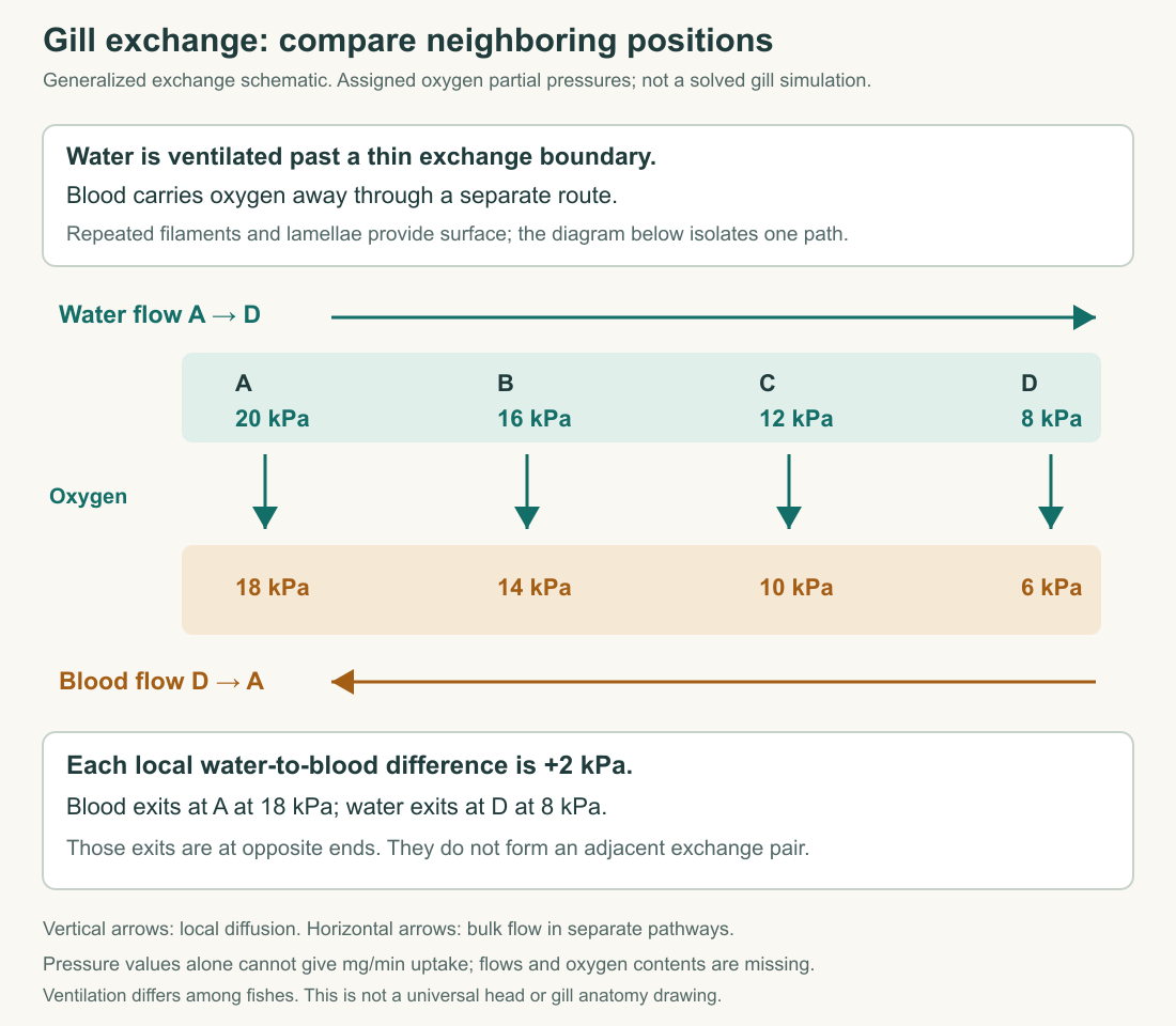
Countercurrent means the two fluids travel in opposite directions along the exchange region. Use four positions, A through D, laid out from left to right. Let water travel A → B → C → D, and blood travel D → C → B → A. The local comparison is between adjacent water and blood at the same position.
Assign the water oxygen partial pressures 20, 16, 12, and 8 kilopascals at A, B, C, and D. Assign the blood values 18, 14, 10, and 6 at those same positions. A kilopascal, abbreviated kPa, is a pressure unit. These are invented teaching values illustrating gradients, not measurements from a fish or a solved quantitative gill model.
At every listed position, water exceeds blood by 2 kPa. Follow a parcel of blood from its entrance at D: its assigned partial pressure rises from 6 to 10, then 14, then 18. At each stage it is beside water with a higher value. The blood can therefore continue gaining oxygen along the route without requiring an uphill local diffusion step.
The exiting blood at A has a value of 18, higher than the exiting water at D with a value of 8. That is not a contradiction. Those exits are at opposite ends and were not the adjacent pair in the last exchange step. The blood's final local partner is the incoming water at A, assigned 20. Comparing opposite exits as though they touch creates the apparent paradox.
Reversing an arrow changes the explanation
Now imagine both fluids entering at A and moving toward D. Water begins with the higher partial pressure and blood with the lower. As exchange proceeds, their values approach one another locally. If they reach the same value, the local net diffusion drive disappears. A passive exchange route cannot simply continue transferring oxygen in the same direction after the local gradient has reversed.
Do not reuse the countercurrent table unchanged after reversing the blood arrow. Doing so would have blood move from 18 toward 6 while the intended explanation says it gains oxygen. A diagram's arrows and values must describe the same trajectory. This is a useful test for any exchange schematic: trace a single parcel in its stated direction and ask whether the labels make sense.
Neither arrangement supplies a universal percentage of oxygen extraction. The outcome also depends on incoming conditions, flow rates, barrier properties, available surface, and blood binding. Countercurrent organization preserves useful local differences; it does not guarantee complete extraction or a particular arterial value in every living fish.
The table deliberately specifies partial pressures only. To calculate oxygen amounts transferred, we would need the relationship between pressure and oxygen content in each fluid, along with their flow rates. Adding the pressure drops as though they were milligrams would mix different quantities. A good explanatory diagram can establish a directional possibility without claiming to solve the full material balance.
Calculate uptake from flow and concentration
For a separate original material account, let two liters of water pass through a defined exchange route each minute. Its entering oxygen concentration is eight milligrams per liter and its leaving concentration five. The difference is three milligrams per liter. Multiplying by two liters per minute gives a net removal of six milligrams per minute under the stated steady-flow assumptions.
The incoming oxygen rate is sixteen milligrams per minute, so removal is 6/16, or 37.5 percent. Now double water flow to four liters per minute while leaving the incoming concentration at eight and assigning the outgoing concentration six. Removal becomes eight milligrams per minute, while the extracted fraction falls to 25 percent. Uptake has increased even though fractional extraction has decreased.
That example explains why one percentage cannot rank the total performance of two conditions. A lower extracted fraction can coexist with a higher uptake rate when more water passes through. The example does not prove a real gill will produce those outgoing concentrations after a flow change. Those values would have to be measured or predicted using a supported model.
The route also needs a clear boundary. If some sampled water bypassed the exchange region, or another process consumed oxygen between the inlet and outlet, the calculated loss would not automatically equal fish uptake. A mass balance is powerful because it states what crosses a boundary, but it still depends on what the apparatus includes.
Blood delivery completes another part of the route
Blood leaving the gills must carry oxygen toward tissues and return after tissue exchange. The rate delivered to a tissue region depends on blood flow and arterial oxygen content. The amount removed by that region depends on the difference between incoming and outgoing oxygen contents. These are content measurements, not merely partial-pressure readings.
Assign a fictional tissue region a blood flow of 100 milliliters per minute. Suppose arterial blood contains 0.10 milligrams of oxygen per milliliter and returning blood contains 0.04. Arterial delivery is ten milligrams per minute; tissue removal is six. The remaining four milligrams per minute travels back in the venous blood. “Deoxygenated” blood need not contain zero oxygen.
The assigned contents are teaching values, not a reference interval for any species. Their purpose is to separate delivery from extraction. Doubling arterial delivery does not prove tissue use doubles, and increasing tissue extraction can change use without the same increase in flow. The complete interpretation needs both sides of the blood route.
This returns us to the working muscle from Chapter 2. More movement can change oxygen demand, but a visible tail beat does not specify gill uptake, blood delivery, or tissue extraction. An explanation should identify the part of the cascade being measured and connect it to the others using explicit evidence or assumptions.
The exchange surface has other obligations
The gill's large exposed interface also participates in water and ion exchange, acid–base regulation, and nitrogen-waste handling. Evans, Piermarini, and Choe's review emphasizes these connected roles. A surface that favors gas exchange can create additional demands for maintaining internal composition, so “make the gill as large and permeable as possible” is not a complete design principle. Evans and colleagues: the multifunctional fish gill
This is where the breathing chapter meets the later salt-balance chapter. Increasing access between body and environment can help one transfer while making another harder to control. The specific transport mechanisms matter; it would be equally misleading to assume that every substance crosses at the same rate simply because the surface is thin.
For now, our complete route is water movement, local diffusion across a living barrier, oxygen binding and transport in blood, then tissue exchange and use. Each step has a location and a rate. The fish can breathe because these processes work together, while differences among fish determine how that cooperation is achieved.
Application
Explain the countercurrent table
Draw the four positions and both flow arrows. Enter the assigned water and blood partial pressures. Trace blood from entry to exit and explain how its exit value can exceed that of the exiting water without any listed uphill diffusion step. State one quantity the diagram cannot calculate on its own.
Diagnose a misleading percentage
Condition one removes six milligrams of oxygen per minute at 37.5 percent extraction. Condition two removes eight at 25 percent. A report calls condition two worse because the percentage fell. Explain what its ranking misses, then name an additional quantity needed to compare the energetic costs of the two conditions.
Model interpretation
Blood moves D → C → B → A through values 6, 10, 14, and 18 kPa. At each location, the adjacent water is 2 kPa higher. The outgoing blood at A meets incoming water at A, not outgoing water at D. Comparing the two exits ignores their locations. The table does not provide oxygen content relationships or flows, so it cannot calculate a transfer rate in milligrams per minute.
The second condition removes more oxygen per minute despite a smaller fraction of the incoming oxygen supply. Its water flow is greater. Comparing cost requires information about the work of moving water and the relevant metabolic expenditure, not extraction percentage alone. Neither the assigned example nor a mouth-opening count supplies that cost.
The fictional blood account likewise separates ten milligrams per minute delivered from six removed. Returning blood still carries oxygen. A complete interpretation labels fluid direction, local gradient, total content, and rate rather than using “more oxygen” for all four.