Looking past the familiar silhouette
A moon jelly passes the aquarium window with its bell facing you. A moment later it turns sideways and seems to become a different animal. The disk becomes a dome. Structures that appeared to lie on its surface now hang beneath it. The apparent disappearance of a body part can be nothing more than a change of view. Before explaining how this animal swims or feeds, we need to make its transparent body intelligible.
Our recurring example is the moon jelly that Monterey Bay Aquarium identifies as Aurelia labiata, a species found in the northeastern Pacific, including Monterey Bay. Its broad bell, short marginal tentacles, and folded oral arms offer an accessible starting point. The aquarium's species account supplies this identity and local setting. When research concerns a different moon jelly, we will name it. “Moon jelly” is a useful common name, but it cannot carry every result obtained from every animal assigned to Aurelia.
The first useful question is surprisingly practical: where is outside? Imagine a drop of seawater directly beneath the dome. Is it inside the jellyfish? Geometrically it lies within the outline of the bell; biologically it can still be outside the animal. Confusing those two meanings of inside produces a mistaken explanation of both feeding and propulsion.
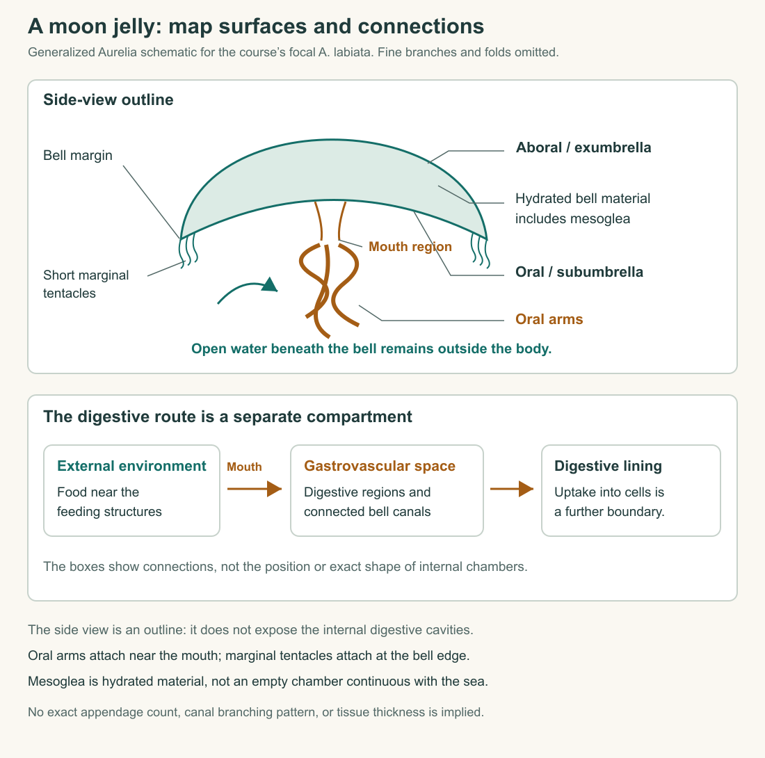
Find an axis before naming the parts
Start with the mouth rather than the direction of travel. The mouth is on the oral side: the side associated with feeding. The opposite surface of the bell is aboral, meaning away from the mouth. These names remain usable when the animal tilts, rolls, or changes its swimming direction. “Top” and “bottom” depend on how it happens to be oriented in the tank. Anatomical language earns its place by staying attached to the animal.
The bell's outer convex surface is the exumbrella; the concave oral surface is the subumbrella. Picture an open umbrella without its handle. Rain meets the convex surface, while your head occupies the space beneath the concave surface. Your head is sheltered by the umbrella, not inside its fabric. Likewise, water under a jellyfish bell need not have entered its digestive system. The analogy explains a boundary, not the material or complete shape of an actual jellyfish.
Now add the feeding structures. Oral arms extend from the mouth region. They are distinct from the much shorter tentacles arranged around a moon jelly's bell margin. A distant image may blur those structures into one fringe, yet location matters: “an appendage” gives us less information than “an oral arm beside the mouth” or “a tentacle at the bell edge.” Later we can follow material between them. For now, preserve the distinction instead of assuming every projection has the same connections.
A view along the oral–aboral axis shows repeated structures arranged around the center. A side view reveals their depth and attachment. Neither view is a complete anatomical map. Consider a drinking glass photographed straight down: the circular rim and base may overlap in the image, although they are far apart vertically. Transparency adds another difficulty. You can see an internal object without knowing whether it touches the near surface, lies in the middle, or sits against the far surface.
Make a three-view sketch before adding detail. In the oral view, draw the bell outline, the central feeding region, and the margin. In the side view, draw the dome, its concave underside, and the hanging arms. In the cross-section, mark actual tissue separately from open water. A cross-section is an imagined slice, not an ordinary photograph. Label it accordingly. Otherwise, an explanatory drawing can accidentally imply that an animal normally has an open wound exposing its interior.
Repeated organization around an axis is often called radial organization. That description does not mean every possible slice is identical. A slice through an oral arm intersects a structure that a slice between arms misses. Try drawing four equally spaced marks around a circle, then turning the page a little: the circle remains unchanged, while the marks move to different positions. Both the continuous outline and the repeated arrangement belong in the account. In an actual medusa, folds, developing canals, and individual variation add further detail. Begin with the organizing pattern, then locate departures from the simplest drawing rather than erasing them.
The tissue is doing work
The bell is not a bag of seawater with some organs floating inside. Cnidarian anatomy distinguishes an outer epithelial layer, an inner digestive lining called the gastrodermis, and intervening material called mesoglea. The OpenStax introduction to cnidarians provides this basic map and the distinction between a swimming medusa and an attached polyp. Here, the crucial point is spatial: tissue layers enclose and organize spaces. A large transparent region is not automatically a hollow chamber.
Mesoglea is highly hydrated, but hydration does not make it mechanically equivalent to water. In work on animals identified as Aurelia aurita, researchers measured the behavior of its extracellular matrix at different scales. The material behaved as a viscoelastic gel in larger-scale tests, while small probes revealed local heterogeneity. The study's abstract and introductory account connect these properties to a fibrous matrix. The useful conclusion is that a transparent, water-rich material can transmit forces and change shape in a structured way.
“Viscoelastic” combines two kinds of behavior. An elastic contribution stores energy during deformation and can return some of it as the material recovers. A viscous contribution dissipates energy as deformation proceeds. Neither description means that the bell is a perfect spring or that its shape recovers without losses. The distinction will matter when we ask how contraction and recovery alternate during swimming.
Use an invented material comparison to sharpen the idea. Suppose two transparent samples both contain a large proportion of water. Under the same small load, sample A changes shape quickly and continues to flow. Sample B deforms, supports the load for a time, and partly recovers when the load is removed. Their appearance and water content have failed to predict the behavior that interests us. We need measurements of deformation, time, and recovery. These assigned samples are a reasoning exercise, not a jellyfish experiment.
The same logic prevents a common error about body size. A thick bell need not put every living cell equally far from a supply of oxygen: much of the visible thickness can be extracellular material. To assess a cell's exchange problem, locate the cell and the relevant surface. Measuring the entire bell thickness and calling that the diffusion distance skips the anatomy we are trying to explain.
Two spaces that a silhouette conceals
Return to the seawater beneath the bell. That water is continuous with the surrounding sea through the broad opening under the dome. It contacts an external surface. Now consider water and food that have passed through the mouth into the gastrovascular cavity. They occupy a different compartment, bounded by digestive tissue. The mouth connects this internal digestive space to the environment; it does not turn all water beneath the bell into stomach contents.
The word gastrovascular joins digestion with distribution. In a moon jelly, the internal system includes central digestive regions and canals extending through the bell. These canals are not miniature vertebrate arteries carrying blood from a heart. They are parts of a connected digestive and distributing system. Calling every branching channel a blood vessel would replace the animal's organization with an imported human diagram.
Research on Aurelia canal development makes that organization especially visible. Investigators followed growing networks in cultured moon jellies identified in their study as Aurelia aurita. New branches and connections varied during growth; proposed mechanical explanations remained hypotheses. The study's observations and anatomical description show why a canal diagram is a map of a connected system, not a promise that every individual has an identical arrangement of fine branches.
Imagine tracing two colored dots on a drawing. The blue dot begins in seawater under the bell and moves out past the margin. It has crossed no mouth or digestive lining. The orange dot represents a food particle delivered through the mouth into a digestive compartment. Its later chemical breakdown and uptake require additional steps; moving it into the cavity has not yet made it part of a living cell. The routes differ even if the dots temporarily occupy nearby positions in a side-view image.
This distinction also helps with magnification. At whole-animal scale, you can follow food toward an opening. At tissue scale, you can ask where digestive products encounter an absorbing surface. At cell scale, you can ask what crosses a membrane. Those are connected questions with different resolutions. A whole-body arrow labeled “nutrition” conceals the transitions; a useful explanation slows down at the boundaries where the mechanism changes.
Resemblance is a starting point for comparison
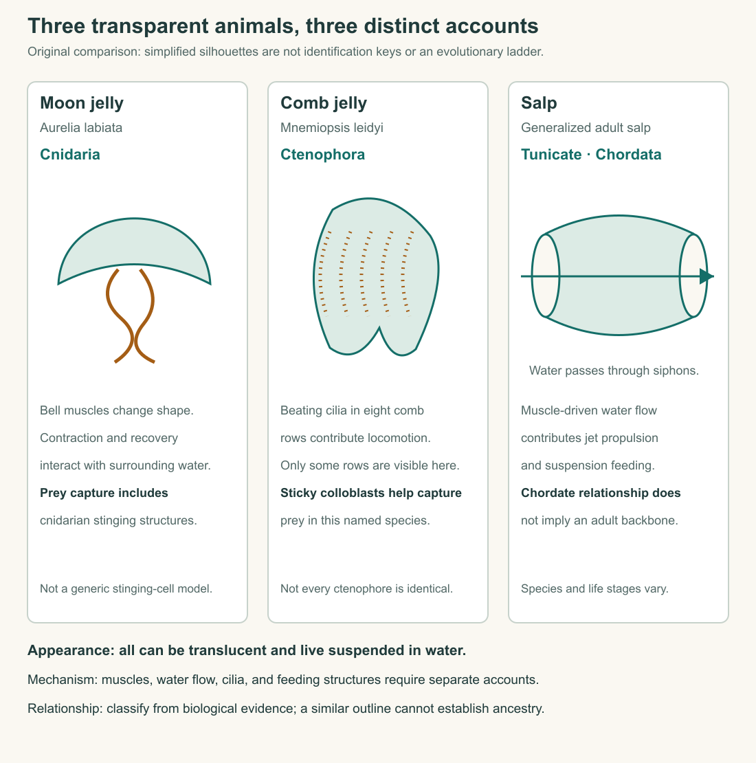
An aquarium may display a moon jelly, a comb jelly, and a salp as translucent drifting animals. That grouping makes sense for a visitor interested in life suspended in water. It does not establish that all three share the same body plan or belong to the same animal lineage. We should compare the feature that interests us, then check relationship separately.
The moon jelly is a cnidarian. Sea anemones and corals also belong to Cnidaria, despite looking very different from a swimming bell. Their inclusion is a useful correction to silhouette-based classification: a fixed, tentacled animal and a swimming medusa can share important inherited organization. A body form is one aspect of an animal, not a substitute for its evolutionary relationships.
For the comb-jelly comparison, use Mnemiopsis leidyi. The Marine Biological Laboratory's research-organism account identifies it as a ctenophore and describes eight rows of beating cilia used in locomotion. Its sticky prey-capture cells, called colloblasts, also differ from cnidarian stinging structures. This is a comparison between specified animals. It does not license giving every ctenophore every feature of Mnemiopsis.
A salp belongs among the tunicates, within the chordate lineage. Woods Hole Oceanographic Institution's account of thaliacean relationships places salps alongside other tunicate groups. A salp's transparent appearance does not move it into Cnidaria. Conversely, its relationship to other chordates does not mean that an adult salp contains a little fish skeleton hidden in its body.
Construct a comparison with three separate columns: observable appearance, operating mechanism, and relationship. “Transparent body” belongs in the first. “Ciliary comb rows” belongs in the second when supported for the animal concerned. “Ctenophore” belongs in the third. Trouble begins when an entry migrates between columns without argument: transparent therefore jellyfish; jellyfish therefore bell propulsion; chordate therefore backbone. A well-designed table stops these shortcuts before they become a narrative.
Identify the animal without pretending the name is effortless
The scientific name itself needs care. A study of Aurelia systematics found extensive overlap and variability in morphological features and emphasized molecular characters in distinguishing species. Its methods and diagnostic discussion make a practical point for this course: a familiar moon-jelly appearance does not always settle species identity. Older research using a broad species name may need to be interpreted in its historical identification context.
That uncertainty does not make anatomy useless. It changes the specificity of the claim. From a clear image, you might confidently describe a bell contracting while remaining unable to determine the species. A labeled aquarium record can support the institution's identification of its focal animal. A carefully identified experimental population can support a species-specific result. Those are different evidential situations, and each can still teach us something.
Suppose an image archive contains three photographs: one frontal view labeled only “moon jelly,” one side view with an aquarium identification, and one microscope image from a documented research population. You want to explain the bell's gross shape. The first two may both help. You want to report a measured property for a species. The attractive frontal photograph adds little unless its identity and measurement context are supplied. Choosing evidence depends on the question, not on which image looks most authoritative.
This is also why the course's drawings are schematics. A schematic can clarify a route by leaving out fine detail, provided it tells you what has been simplified. An anatomical illustration becomes misleading when simplified branches look like an exact count, when tissues become empty spaces, or when arrows suggest a measured speed that was never measured. Read the caption as part of the argument.
You can now look through the aquarium glass with a more useful set of questions. Which surface is facing you? Is a visible space external water or an internal digestive compartment? Which structures connect to the feeding region? What is the animal's identity, and how securely is it known? These questions turn a translucent silhouette into a body whose operation can be investigated. Next, we will put the body back into motion and ask what one pulse actually accomplishes.
Application
Draw a generalized Aurelia medusa in side view and oral view. Label the mouth, oral arms, marginal tentacles, outer bell surface, concave bell surface, mesoglea, and gastrovascular space. Add one arrow showing seawater moving through the open region beneath the bell and another showing a food particle entering through the mouth. Use a different line style for structures visible through tissue.
Check your drawing: the seawater arrow must not enter the digestive cavity merely because it passes under the dome. Mesoglea must appear as material between tissue layers, not as the same empty space as the cavity. Oral arms and marginal tentacles should attach in different regions. The two views must describe the same animal orientation and connections, even though their outlines differ.
Then correct this museum label: “All three are jellyfish: the moon jelly, Mnemiopsis, and a salp have transparent bodies, so they are closely related and swim the same way.”
Model correction: the shared appearance makes a useful ecological comparison but does not establish close relationship or identical propulsion. The moon jelly is a cnidarian; Mnemiopsis is a ctenophore with ciliary comb rows; the salp is a tunicate in the chordate lineage. A separate account of movement is needed for each. An adequate correction keeps the useful comparison while removing the unsupported inference.