Skin as an active surface
An animal's appearance can change even when the pigment molecules in its skin have not been replaced. A colored area may expand; a reflecting structure may redirect light; the skin's surface may rise into a different relief. A camera records the combined result. To explain it, we must separate the tissues producing the appearance from the conditions under which another animal sees it.
This chapter uses European cuttlefish, Sepia officinalis, for two experiments, and longfin inshore squid, Doryteuthis pealeii, for a cellular study. These are deliberate comparisons. Neither is the California market squid, and neither is an octopus. The differences are part of the lesson rather than exceptions to be forgotten once the photographs become striking.
Expand a pigment-bearing surface
A cephalopod chromatophore organ includes a pigment-containing cell and associated radial muscles and nerves. When the muscles contract, they spread the pigment-bearing structure into a larger visible area. Relaxation permits it to retract. A rapid change in displayed area does not require synthesizing a new batch of pigment at the same speed. The system alters the presentation of existing material.
In a 2019 study of D. pealeii, researchers combined microscopy and biochemical analysis to examine pigmentary and structural effects within chromatophore organs. Their findings caution against treating pigment and reflection as perfectly isolated layers with no interaction. The simplified diagram below shows major functional distinctions, not an exhaustive skin section or a claim that every cephalopod has the same arrangement. Dynamic pigmentary and structural coloration.
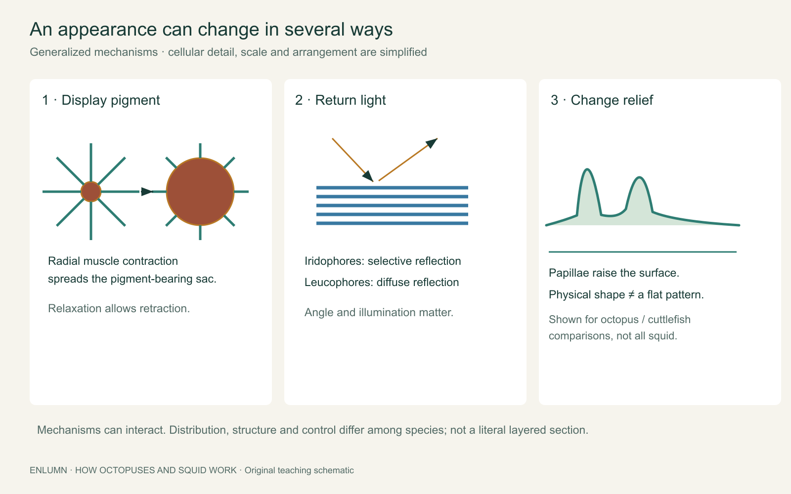
Think of a colored spot whose radius changes from one arbitrary unit to two. If it is approximated as a flat circle, its displayed area becomes four times as large. It has not acquired four times as much pigment. Instead, the same pigment-bearing material can occupy a different shape and thickness. This is an invented geometry exercise, not a measured expansion ratio.
Now place many such elements in a patch. Expanding every element equally may change average appearance, but a pattern requires differences across locations. A dark region next to a pale region cannot be explained merely by saying “the chromatophores expanded.” Which elements changed, by how much and in what spatial arrangement? The scale of the pattern is a separate variable from the state of any one element.
That distinction matters when examining a magnified view. A close-up may reveal individual elements but omit the arrangement a predator sees at a distance. A distant image may reveal the body pattern while blending the smaller structures together. Neither scale is sufficient for every question. Useful analysis connects them while preserving the limits of the image.
Reflection is different from pigment absorption
Pigments alter appearance by absorbing some wavelengths more than others. Structural coloration depends on the way organized material interacts with light. Iridophores can produce selective reflections; leucophores produce more diffuse pale reflections. Their distribution and properties vary. These terms identify optical mechanisms, not a universal set of buttons available to every species.
A reflected color can depend on viewing angle and illumination. If an observer moves, a patch may appear to change without the animal changing the patch's state. Conversely, a biological change can alter the light returned to a stationary observer. The visible outcome alone does not tell us which variable changed. Recording the light source and camera position is therefore relevant biological information.
Try an imagined two-image comparison. In the first image, a patch is darker and the entire background is also darker. In the second, both are brighter. One explanation is altered exposure or illumination. If a fixed reference patch changes by a similar amount, that explanation gains support. If the reference remains stable while a bounded region of skin changes, a local biological change becomes more plausible. This is a comparison design, not proof from a hypothetical example.
Camera settings can conceal real change as well as invent apparent change. Automatic exposure may compensate when the animal darkens, making the surrounding water brighter. White balance can shift relative colors. An image analysis that ignores these adjustments can mistake an instrument's response for the animal's response. The measurement process belongs in the explanation.
No single photograph establishes how well a pattern conceals an animal from all predators. Different visual systems sample light differently, and viewing distance changes which details remain resolvable. A pattern that appears conspicuous in a tightly cropped, brightly lit image may be less detectable in a wider scene. The relevant observer and conditions must be specified before camouflage success can be evaluated.
Distance introduces another measurement choice. If two small spots merge into one blurred patch in a photograph, their apparent number falls although neither disappeared. A comparison should preserve scale or explicitly account for the change in resolution. Enlarging the final image afterward cannot recover detail the original recording failed to resolve. Thus, image size on a screen is not the same thing as the biological scale or resolving power of the observation.
Texture can mean two different things
A spotted pattern is sometimes called texture even if the surface is physically smooth. A raised bump is also texture, but it changes three-dimensional form. Papillae are projections of the skin that can alter relief in species that possess suitable structures. Distinguishing pattern from relief prevents a misleading explanation in which changing color is assumed to produce every visible bump.
A paper model makes the distinction concrete. Draw dark triangles on a flat sheet, then fold a second sheet into ridges without coloring it. Both can create visual edges, but one changes pigment arrangement and the other changes geometry. Lighting from the side may make the ridges conspicuous through shadows. Diffuse lighting may reduce that effect. A real skin surface can combine these mechanisms.
Allen and colleagues asked whether European cuttlefish required tactile information from a substrate to express selected papillae. Their 2009 comparison included actual substrates, substrates under glass and laminated photographs. The reported responses supported control by visual cues under those conditions, while some papilla groups showed irregular responses that required further investigation. The study's abstract supports this bounded conclusion; it does not establish that every skin response in every cephalopod is controlled in the same way.
The experimental logic is accessible without pretending to repeat the experiment. Covering a substrate with glass changes access to its physical texture while preserving substantial visual information. A photograph removes direct contact with the original relief and also changes other aspects of the scene. Comparing several versions helps test an explanation, but none is a perfect copy differing in only one imaginable variable.
Suppose you wanted to strengthen the inference on paper. You would ask how illumination, reflections from glass, image scale and the animal's position were controlled. You would specify which papillae were scored rather than averaging all surface change into one number. You would also distinguish no detected difference from proof of exact equality. Each improvement makes the proposed mechanism more testable without requiring a grander claim.
A pattern transition need not follow a fixed script
The familiar labels uniform, mottled and disruptive can help describe appearances. They do not require the nervous system to select from only three stored pictures. Woo and colleagues' 2023 study tracked European cuttlefish over changing backgrounds using images at different scales. Transitions through measured pattern space often followed variable, meandering paths rather than a single stereotyped route. The study concerned two-dimensional skin patterns, not a complete measurement of every camouflage mechanism. The dynamics of pattern matching.
To understand pattern space, imagine representing each image by two invented measurements: the fraction of a patch that is dark and the size of its largest dark region. Each image becomes a point on a graph. A sequence of images becomes a path. The graph is not the physical route traveled by the cuttlefish; it is a representation of changing appearance.
Consider points A = (2, 2) and B = (8, 8). A direct path could pass through (4, 4) and (6, 6). Another could pass through (6, 2), then (4, 7), then B. Both reach the same final point, but their trajectories differ. Endpoints alone would hide that difference. The invented coordinates are teaching devices, not data extracted from the study.
Now consider the opposite limitation: two visually different patterns may share the same two measurements. A stripe and a cluster of spots can have equal dark area and an equally large region under a coarse definition. The representation has discarded other features. A two-dimensional plot therefore cannot automatically show the full complexity of the underlying patterns. Choosing measurements is part of the scientific work.
Likewise, a meandering trajectory is evidence about the process, but it does not by itself identify every feedback pathway or prove that the animal consciously evaluates each alternative. Mechanistic proposals must connect the trajectory to information, neural activity and control. Describing a pattern as a search can be useful mathematically while remaining silent about the animal's subjective experience.
Camouflage and communication ask different questions
Camouflage concerns how an appearance affects detection, recognition or targeting by an observer. Communication concerns information conveyed between organisms through a signal and its effects on a receiver. The same kind of skin machinery can participate in different contexts. A bright change during an encounter is not automatically camouflage, and a conspicuous pattern is not automatically a message with a human-like meaning.
Begin with the setting. Was a potential predator present? Was another member of the species nearby? What did the receiver do before and after the change? Was the pattern directed toward a particular observer or associated with a broader change in activity? These observations help frame alternatives; they do not settle them without appropriate comparisons.
Imagine an animal darkening as another approaches. One interpretation is a display affecting the approaching animal. Another is a response to a changed background or illumination. A third is a correlated response to arousal whose communicative role has not been established. Repeated timing and a receiver's reaction can be informative, but shared responses to the same external event remain a possible confound.
A useful paper test would compare receiver behavior when the candidate visual feature varies while other relevant cues remain similar. The feature must be something the receiver can perceive under the conditions. A manipulation that looks obvious to a human camera may be ineffective or unnatural for the animal. We are specifying evidence needed for a claim, not proposing a home experiment with captive cephalopods.
Avoid translating a pattern into a sentence such as “I am angry” unless the evidence supports the underlying behavioral interpretation—and even then the sentence is a metaphor. An observable change in approach or withdrawal is a firmer description than an assigned emotion. We can respect the complexity of an animal's behavior without pretending to possess its internal vocabulary.
Build a fair comparison of two images
Suppose you are given before-and-after images of a cuttlefish. Start by verifying that they show the same individual and identifying whether the interval is known. Record the background, viewpoint, lighting and the region being compared. Then separate changes in pattern, average brightness, reflection, relief and posture. Several may occur together; naming them separately makes their possible causes clearer.
Next, decide what conclusion the images could support. They might show that a particular patch changed appearance. They might suggest that a pattern became more similar to a background under a chosen measurement. They cannot alone establish that predators failed to detect the animal, that a receiver understood a signal or that the same response occurs across all species.
A strong comparison contains a small, defensible claim and a useful next question. For example: “The large pale region becomes less distinct relative to neighboring dark areas in these views. Fixed-reference measurements are needed to separate local change from exposure differences.” That is more informative than “the cuttlefish perfectly copied the sand,” which assumes both mechanism and success without measuring either.
The active skin is remarkable because anatomy, optics and control meet at a surface. Muscle changes the presentation of pigment; reflecting structures affect returned light; relief interacts with illumination; spatial coordination produces a body pattern. The resulting appearance belongs to a relationship between animal, environment and observer. Understanding that relationship prepares us to examine the nervous system without reducing it to either a simple switchboard or a human mind in an unfamiliar body.
Check your understanding: Two images show a cephalopod becoming brighter, but the rock beside it also brightens. What should you check before concluding that its chromatophores retracted?
Expected answer: Check lighting, exposure, viewpoint and a stable reference, then distinguish local skin change from a scene-wide change. Brightness alone does not identify chromatophore state or exclude reflective and geometric effects.
Application
Using the original skin diagram, write separate explanations for a larger colored patch, an angle-dependent reflection and a raised papilla. State which observation would help distinguish them.
Then read the linked papilla-study abstract and the camouflage-study abstract. In 200 words, compare the questions they ask and the measurements needed. Name Sepia officinalis and preserve the difference between physical relief and two-dimensional pattern. Allow 10–15 minutes. No live-animal manipulation is required.