A map that does not become a myth
A cup sits near the edge of a table. You recognize it, decide to move it, reach, adjust your fingers to its shape and lift without spilling. The action feels like one event. It depends on information arriving from several sources, activity distributed through many neural pathways, and muscles working within a changing physical situation. No single colored patch on a brain diagram contains the whole explanation.
This course follows that ordinary task as a way into the human brain. We will examine cells and signals, perception and movement, bodily regulation, learning and the methods used to study them. The aim is to understand an organ well enough to connect its parts without turning a map into a collection of myths.
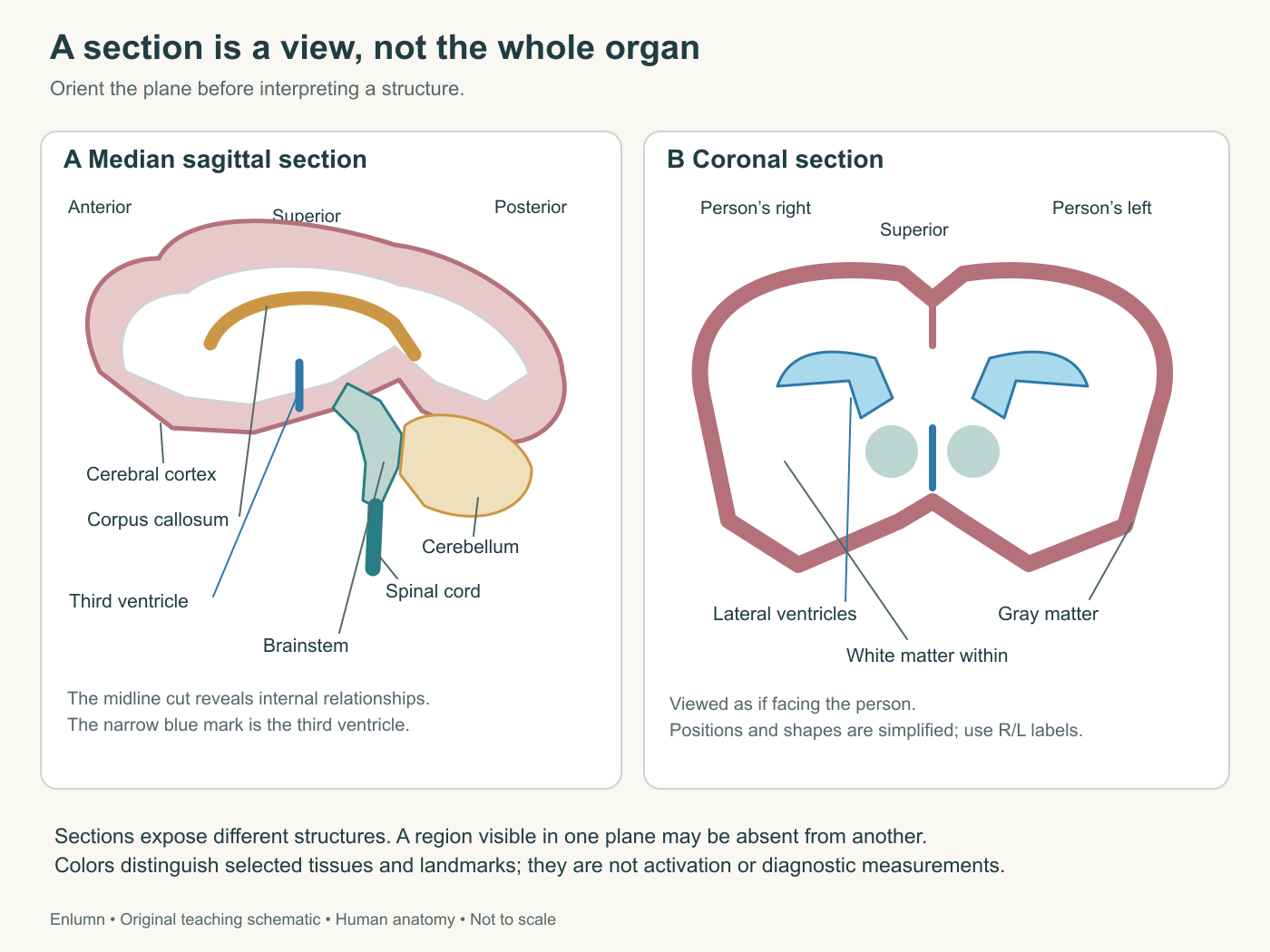
Read the orientation labels before naming a structure. The two panels are different section planes, not successive stages of a process. Their colors identify selected landmarks and tissue categories; they do not show activity. The drawings simplify shape and omit many structures so that the relationship between a section and the whole organ remains visible.
Establish the view before naming the part
Use the person's anatomical directions, not the reader's position on the page. In a front-facing view, the person's right appears on your left. A side view exposes one lateral surface; a view toward the midline reveals a medial surface. The shared human-anatomy orientation introduces these conventions along with tissues, flow and feedback. Revisit it when needed rather than memorizing a separate orientation for every organ.
Anterior means toward the front and posterior toward the back. Superior means above and inferior below in anatomical position. Medial means toward the midline, while lateral means away from it. These are relationships. A point can be medial to one structure and lateral to another without contradiction. Include the reference when a description might otherwise be ambiguous.
Three common section planes expose different relationships. A sagittal section divides right from left; a midsagittal section passes through the midline. A coronal section divides front from back. An axial, or transverse, section divides upper from lower regions. Real imaging can use tilted planes, so these terms describe orientation rather than guaranteeing that every image follows an ideal textbook slice.
Imagine cutting a curved tube at different locations and angles. A cross-section can look round, oval or elongated, while a section along its length may reveal a continuous passage. The object did not change between views. The section plane changed what became visible. This simple geometrical problem explains why a brain structure can appear in several separated profiles across an image series.
Before interpreting an unlabeled scan, ask where its right and left markers are and how the image is displayed. Radiological display conventions can differ from the intuitive view a beginner expects. You do not need to diagnose scans in this course, but you do need to recognize that orientation must come from the image's information. Guessing from a familiar silhouette is an unreliable substitute.
The folded surface is only part of the cerebrum
The cerebrum consists of two large hemispheres and their internal structures. Its outer cerebral cortex is a sheet of gray matter folded into ridges, or gyri, and grooves, or sulci. The surface is striking, but it is not the entire cerebrum and certainly not the entire brain. Beneath it lie pathways and deeper structures that participate in the same activities.
The major surface lobes—frontal, parietal, temporal and occipital—are useful geographical divisions. The central sulcus separates frontal and parietal regions; the lateral sulcus marks an important boundary above the temporal lobe. Some boundaries are more evident from one surface than another. The insula lies deeper within the lateral fissure and is hidden in an ordinary external view. A map of four visible lobes is therefore a useful beginning rather than a complete inventory.
The OpenStax central nervous system section supplies anatomical orientation for these major regions. Use its diagrams to locate structures, while resisting the temptation to read a short functional label as an exclusive assignment. We will develop the connections and qualifications that a small map cannot contain.
Gray matter contains concentrations of neuronal cell bodies, dendrites, synapses and other tissue components. White matter contains many myelinated axons, alongside glia, vessels and other structures. Neither is made of only one cell type. White matter is not empty cable insulation, and gray matter is not the only place where living processes occur. Their difference describes organization and appearance, not a division into important and unimportant tissue.
A nucleus, in this anatomical context, is a grouping of neuronal cell bodies within the central nervous system. It is not the same use of the word as a cell nucleus. A tract is a bundle of axons within the central nervous system. These terms let us distinguish a region containing many cell bodies from a route containing many projections, while remembering that real boundaries and connections can be complex.
The corpus callosum is a major white-matter connection between the cerebral hemispheres. Its presence immediately complicates the popular image of two independent brains, one logical and one creative. Some functions are lateralized, meaning their contributions are not perfectly symmetrical, but asymmetry does not establish two types of person. A task can draw on both hemispheres while depending more heavily on particular operations on one side.
Look beneath the cortex
The thalamus is a collection of nuclei in a central location with extensive connections to cortex and other structures. It participates in the routing and processing of information rather than acting as an inert relay box. The visual pathway we will follow includes a thalamic stage, but different thalamic nuclei have different connections. “The thalamus sends the senses to the brain” is misleading because the thalamus is already part of the brain and the pathways are not all identical.
The hypothalamus lies below the thalamus and participates in regulation of the body's internal state, including interactions with autonomic and endocrine systems. Its position near the pituitary helps orient a diagram, but location does not substitute for a physiological account. A regulatory effect may involve a chain of neural and hormonal signals extending well beyond the structure where the diagram places its label.
The basal ganglia are interconnected structures involved in circuits supporting movement, learning and other functions. The conventional name is retained even though ganglion usually refers to a grouping outside the central nervous system. Their connections with cortex, thalamus and brainstem matter more for our purposes than memorizing an isolated list. In the movement chapter, we will distinguish selecting and modulating actions from directly contracting a muscle.
The hippocampus lies in the medial temporal region and is important in forms of learning and memory. The amygdala, nearby but distinct, participates in processing biologically significant information and in wider systems associated with emotion and learning. Calling one a memory storage box and the other a fear button turns useful associations into false boundaries. Different kinds of memory and emotion involve different combinations of processes.
These deeper structures are easily omitted from a side-view sketch because they are not visible on the outer surface. Their absence from that sketch is not evidence of lesser importance. A surface map and a section map answer different questions. Use both before deciding that a behavior belongs entirely to the brightly colored lobe you can see.
The brainstem and cerebellum are not optional additions
The brainstem includes the midbrain, pons and medulla. It connects with the spinal cord and contains pathways and nuclei involved in movement, sensation, arousal and bodily regulation. Some cranial nerves connect with it. The spinal cord is part of the central nervous system, but it is not merely a passive extension cable: it contains circuits that integrate information and organize responses.
The cerebellum lies behind the brainstem and beneath the posterior cerebrum. It has a folded cortex of its own, white-matter pathways and deep nuclei. It contributes to coordination and adaptation through its connections with other regions. “Balance center” captures only a small part of its relevance, while “muscle controller” can imply a direct route to muscles that the simplified account does not establish.
For the cup-reaching task, notice how many different kinds of information can matter. You need to estimate the cup's location, relate it to the hand, generate an appropriate movement and adjust when contact supplies new information. A system that compares expected and incoming consequences can contribute to correction and learning. That is a functional question to pursue through pathways and evidence, not a reason to assign the whole task to the cerebellum.
The distinction between a structure and a process is fundamental. A region can contribute to several processes, and a process can require several regions. This many-to-many relationship is not an excuse for saying that every part does everything. Anatomical and experimental evidence constrain the contributions. The challenge is to be specific without pretending that a complex activity occupies one isolated address.
Coverings and fluid are part of the working environment
The brain is soft tissue enclosed within the skull. Three connective-tissue coverings, the meninges, surround the central nervous system. From outside inward, they are dura mater, arachnoid mater and pia mater. The pia closely follows the surface; the arachnoid bridges over it, leaving a subarachnoid space that contains cerebrospinal fluid. A covering, a space and a fluid must remain separate labels.
Cerebrospinal fluid, or CSF, occupies connected spaces within and around the central nervous system. The internal spaces include paired lateral ventricles, the third ventricle and the fourth ventricle. Choroid plexuses contribute to its production through regulated transport. The circulation and coverings reference gives the basic ventricular arrangement. CSF is not simply whole blood squeezed through a sieve, and its routes are not the same as the routes of blood perfusing brain tissue.
A broad pathway leads from the lateral ventricles into the third, through the cerebral aqueduct to the fourth, then into spaces around the brain and spinal cord through openings. This is a useful route for orientation, not a claim that every molecule follows a single unbranched conveyor belt. Production, exchange and drainage occur in a living system with multiple interacting routes.
The brain's arterial supply arrives through internal carotid and vertebral systems. Blood passes through vessels and exchange regions before returning through veins and venous sinuses. Connections among arterial territories can provide alternative routes under some conditions, but they do not guarantee uninterrupted supply if a vessel is obstructed. A diagram of connected vessels is not a promise that every connection can deliver enough flow.
This distinction parallels the cup task. An anatomical route supplies a possibility; its actual performance depends on conditions. A narrow connection, inadequate pressure difference or altered tissue can limit flow despite a visible line on the map. Physiology explains what happens through the structure. Anatomy tells us which relationships that explanation must respect.
Practice a network explanation without erasing location
Draw five boxes labeled visual input, object and spatial processing, action selection, motor output and sensory feedback. Connect them into a loop rather than a one-way chain. This is a task model, not an anatomical diagram. It separates operations that can overlap in time and depend on multiple pathways. The loop reminds us that reaching changes what is seen and felt, which can change the ongoing action.
Now add candidate anatomical regions beside the relevant operations, using cautious wording such as “contributes through connections with.” The visual pathway includes retinal and thalamic stages before and alongside cortical processing. Motor output ultimately reaches spinal motor neurons and muscles. The boxes should not suggest that the cup's meaning is stored entirely in one region or that every signal must pass through the same sequence.
The purpose of the drawing is to generate questions that evidence can answer. If a person can recognize the cup but reaches inaccurately, which operations might be affected? If strength is adequate but an intended action is difficult to initiate, what different processes might be relevant? These are fictional contrasts for understanding, not enough information to diagnose a real person or localize an injury.
A network can be disrupted at a node or along a connection. Removing one bridge from a transport network may affect distant destinations that remain physically intact. Similarly, a brain lesion can alter the functioning of connected regions. An observed deficit does not necessarily mean that the damaged tissue alone contained the missing ability in a compact, complete form.
Yet distributed organization does not make anatomy irrelevant. A damaged pathway can have a characteristic effect precisely because its connections are specific. The appropriate conclusion often lies between two extremes: a solitary center that does everything and a homogeneous brain in which every location is equivalent. A useful explanation names a contribution, a connection and the evidence for both.
Read a brain map as a set of claims
When you encounter a colored brain image, identify what the colors represent. They may distinguish anatomical regions, tissue types, a measured signal or a statistical comparison. A colored region in an illustration is not necessarily a region observed to be active. A region absent from a thresholded activity map is not necessarily inactive. The legend and method determine what can be inferred.
Also ask whether the image represents one person, an average or a schematic. Averaging can clarify a shared pattern while smoothing individual differences. A schematic can clarify a relationship by distorting proportions. Neither is inherently deceptive when labeled honestly. Problems arise when the precision of an image is mistaken for precision in the claim it supports.
Your first map should remain deliberately revisable. It should distinguish hemispheres, surface and deeper structures, brainstem, cerebellum, ventricles and coverings. It should show where the spinal cord connects and where the perspective changes. It should leave space for pathways rather than filling every region with a psychological label.
The next chapter moves from this organ-level geography to the living cells that make it possible. Neurons need gradients, nutrients and a regulated environment; glia have active roles in maintaining and modifying that environment. A map of regions becomes an explanation only when we can follow processes across these scales, from a cell membrane to the coordinated act of moving a cup.
Check your understanding: Why does a labeled “memory region” on a brain diagram fail to show that all memories are stored there?
Expected answer: The label may indicate an important contribution to particular memory processes, not exclusive storage of every kind of memory. Different tasks and stages involve connected regions, and the diagram's evidence and measurement method must be identified before making a broader claim.
Application
Make a side-view and a section-view sketch. Label the cerebrum, cerebellum, brainstem, thalamic region and ventricular spaces where the view permits. Mark right/left or anterior/posterior as appropriate. Use the linked anatomical diagrams to check relationships, not to trace an unlabeled silhouette from memory.
Below the sketches, draw a five-operation model of seeing and moving a cup. Add two feedback arrows and one question about a connection. Allow 15–20 minutes. The drawings are learning tools, not clinical maps or interpretations of personal scans.