Finding the heart in the chest
Put a familiar heart diagram beside a drawing of a chest. The diagram offers four tidy rooms, two above and two below, with a pointed bottom. The chest drawing gives you ribs, lungs, a breastbone, and a diaphragm. Fitting the first picture into the second is less straightforward than it looks. Which chambers face forward? Does the point face straight down? What has the illustrator removed to let you see inside?
These are physiological questions as well as drawing questions. A pump works through connections between spaces, walls, and openings. If we mistake an external covering for a chamber wall, or an overlapping vessel for a connection, the later explanation of blood flow will fail. We will begin by locating the heart as a three-dimensional organ. Its operation will become easier to understand once the flat symbol has stopped dictating the anatomy.
Shared orientation: five tools for the organ courses
Anatomical directions belong to the person being described. In the reference position, the person stands upright with face and palms forward. Anterior means toward the front; posterior, toward the back. Superior points toward the head, inferior toward the feet. Medial means toward the body's midline, lateral away from it. Right and left are the person's right and left, even when you face them. These conventions, introduced in OpenStax's anatomical terminology, remain attached to the body when it lies down.
A plane specifies a possible slice. A transverse plane separates superior and inferior portions; a frontal plane separates anterior and posterior portions; a sagittal plane separates right and left portions. Naming the plane does not tell you which side of the slice you are viewing. A drawing needs direction labels as well. A photograph of a model, a cut surface, and an image assembled from a scan are different representations of the same spatial problem.
A tissue is an organized association of cells and their surrounding material. An organ combines tissues into a working structure. The broad tissue categories are epithelial, connective, muscle, and nervous: surfaces and linings, support and integration, force production, and signaling. The OpenStax tissue account supplies this starting classification. It does not mean an organ has four neatly separated blocks. In a heart wall, vessels, supporting material, muscle cells, and nerve fibers occupy connected arrangements at different scales.
Blood flow and diffusion are different kinds of movement. Flow carries fluid through a route. Diffusion describes molecular spreading, with a net movement of a freely diffusing substance down its concentration gradient under the simplified conditions we will initially use. Crossing a cell membrane also depends on what the membrane permits. The membrane reference introduces these local boundaries. Carrying oxygen near a tissue and getting oxygen into its cells are connected tasks, not interchangeable descriptions.
Finally, regulation needs a loop. In negative feedback, a sensed change produces a response that tends to oppose that change. The general feedback account distinguishes sensing, coordination, and response. A regulated quantity can still vary; opposing a disturbance does not guarantee perfect correction. Later chapters will identify the actual signals and responding structures. For now, a useful check is to ask which variable is sensed and how the response affects that same variable.
Keep these five tools together: directions, sections, tissues, transport, and feedback. They will recur in the organ series, with additional detail introduced where it earns its place. No previous animal-biology course is required.
Place the heart among its neighbors
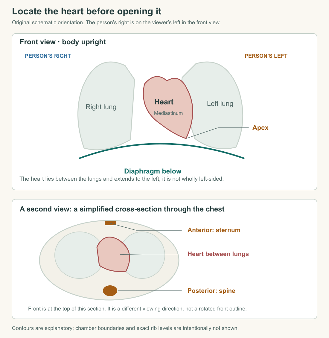
The heart occupies the mediastinum, the central region of the thorax between the lungs. The sternum, or breastbone, is anterior to it; the vertebral column lies posteriorly. Its inferior aspect relates to the diaphragm, the muscular partition between thorax and abdomen. The heart extends across the midline, with much of it toward the left. Saying simply “the heart is on the left” loses the central location and its relationships to both lungs.
The pointed apex is directed inferiorly and toward the person's left. The organ's long axis therefore differs from the body's upright axis. The University of Minnesota's anterior-orientation account makes this relationship explicit. The point of the drawing should help you locate a sloping organ, not persuade you that it normally balances on a vertical tip.
Build an original paper map in three passes. First draw the chest midline and label the person's right and left. Then place the two lungs on either side of a central region. Finally add the heart within that region, extending leftward and resting above the diaphragm. This is a location sketch, not a scale model. Its success depends on the relationships remaining intelligible when the page is turned.
Now imagine the person lies face upward. The heart has not acquired new anterior and posterior surfaces merely because gravity points differently relative to the chest. Anatomical direction and gravitational direction are separate descriptions. This distinction will matter when reading any organ image taken with a person lying down. The labels preserve the body map while the position of the body changes.
Rotate the organ without renaming the chambers
The four chambers are the right atrium, right ventricle, left atrium, and left ventricle. An atrium receives returning blood and communicates with its ventricle; a ventricle ejects blood into an outflow vessel. The NHLBI chamber account also identifies the tissue partitions separating right and left. The singular is septum, the plural septa. A partition is not a fifth chamber or a valve that normally opens once per beat.
The names “right” and “left” do not describe two identical boxes placed cleanly beside each other. Much of the right ventricle lies anterior to the left ventricle. From an external anterior view, the right-sided chambers are more prominent and most of the left atrium is concealed posteriorly. The University of Minnesota's discussion of heart orientation explains how an upright display convention can obscure these relationships. A picture showing all four interiors has made deliberate choices about rotation and exposure.
Try this connection puzzle without drawing a realistic organ. Label four boxes RA, RV, LA, and LV. Connect RA to RV and LA to LV. Place RV partly in front of LV and LA behind them. The connections do not change merely because the boxes overlap in your view. Then spread the boxes apart so every connection is visible. You have improved the route diagram while making it less faithful to position. Both drawings can be useful if their purposes are stated.
The chamber pages for the right ventricle and left ventricle provide a further check: relative position and pumping destination are different facts about the same chamber. A label can tell you where a structure sits, what it connects to, or what it does. A good explanation joins those facts instead of expecting one label to carry all three.
Find the openings, then distinguish them from overlaps
The superior and inferior venae cavae enter the right atrium. Their names distinguish major returning routes from the upper and lower regions of the body. The right ventricle leads into the pulmonary trunk, which branches toward the lungs. On the other side, pulmonary veins enter the left atrium, and the left ventricle leads into the aorta. The left-atrium account is a useful example of identifying a chamber by its inlet and outlet rather than its location on a page.
For the moment, learn this as a connection map. The complete journey through the lungs and body belongs to the next chapter. A vessel drawn behind another vessel has not necessarily joined it. A line passing through an outlined chamber may represent a structure seen in projection, not an open tunnel through its cavity. Transparent drawings need especially clear conventions because they let you see through boundaries that remain physically present.
Give every proposed connection a test: can you trace a continuous lumen from one space to the next? Lumen means the interior space of a hollow structure. A valve can occupy an opening between spaces while regulating passage through it. Tissue separating two neighboring spaces is different. If a diagram shows an uninterrupted wall between two chambers, do not send a flow arrow across it simply because that is the shortest route on the page.
This test also improves a hand-drawn schematic. Leave a small break in the wall where an opening actually exists, label the valve or vessel, and keep other boundaries continuous. If two routes cross without joining, make the separation explicit. These drawing choices are claims about anatomy. They are more consequential than whether the outline looks pleasingly like a heart.
Separate the working wall from the surrounding sac
From a chamber's blood-filled interior outward, the heart wall includes endocardium, myocardium, and epicardium. The endocardium lines the inner surface. The myocardium contains the muscle responsible for the chamber's mechanical work, supported by connective tissue and supplied by vessels. The epicardium is the outer layer of the wall and includes the visceral serous covering. The OpenStax wall description checks this order. “The heart is muscle” identifies a dominant component, not everything composing the organ.
The pericardium is the surrounding sac. Its fibrous component supplies support; its serous component has a parietal layer lining the sac and a visceral layer associated with the heart surface. Between the opposing serous surfaces is a potential space containing a thin film of fluid. The Michigan anatomy account distinguishes these layers and the lubricated interface. A potential space is not normally a large empty chamber waiting to be filled with blood.
Draw two boundaries rather than one thick outline: a working organ wall and an enclosing sac. Magnify the narrow interface enough to label it, but state that the gap is exaggerated. Otherwise, a teaching enlargement can become a false anatomical claim. The blood within a ventricle and fluid at the pericardial interface occupy different spaces with different connections.
A simple mechanical analogy helps locate the question. Imagine a flexible pouch moving inside a supporting sleeve. The pouch material, its contents, the sleeve, and their interface are separate parts of the arrangement. Changing the sleeve's freedom to move could change how the pouch fills even if the pouch wall itself is unchanged. This is a model of interacting boundaries, not a clinical diagnosis or an exact representation of pericardial mechanics.
Blood inside the pump is not the pump's entire blood supply
The heart's muscle cells need their own delivery of oxygen and nutrients. Coronary arteries arise from the aortic root and supply the heart wall through a branching circulation. Returning cardiac veins carry blood away from that tissue. The NHLBI coronary-circulation passage distinguishes this supply from the large streams passing through the chambers. A ventricle full of blood does not eliminate the need to perfuse its working wall.
Make two routes on your sketch. One arrow remains inside a chamber and points toward its outlet. Another follows a vessel supplying the wall and then a smaller exchange region near cells. These routes answer different questions: where does the pump send its contents, and how are the cells doing the pumping supported? The muscle and the circulating blood are coupled without being the same compartment.
This is our first reason to keep several scales visible. A chest diagram locates an organ. A chamber diagram locates a lumen and its openings. A tissue diagram locates vessels, cells, and exchange boundaries. Zooming in does not invalidate the larger picture; it supplies the connections that the larger picture leaves unresolved. Later, a failure of local supply will test whether we have preserved those connections.
Read a section as evidence of a three-dimensional structure
A slice is selective. Cutting a drinking straw straight across gives a circular opening; an oblique cut gives an elongated opening. The straw has not changed into a wider tube. The cut has changed. This original spatial example explains why a two-dimensional chamber outline cannot automatically be treated as the chamber's full size or shape. Orientation and the level of the section matter.
Now imagine a sequence of parallel slices through a model heart. A chamber can enter the sequence, become wider, narrow, and disappear. Its disappearance from one slice does not mean the chamber has ended everywhere in space; the next plane may simply miss it. To reconstruct a route, follow adjacent sections and the vessel's continuity. One attractive cross-section seldom contains every connection needed for a whole-organ account.
The same caution applies to a removed wall. If an illustrator cuts away the front of a ventricle, the newly visible interior is not an external surface. Label the cut edge and name the view. An anatomical drawing should let a reader distinguish what remains in the body, what was removed to expose it, and what was enlarged to make a relationship legible.
You now have enough structure to make the next question precise. Blood returns through particular vessels, occupies particular chambers, and leaves through particular outlets. Those spaces are bounded by a living wall, supplied by another circulation, and situated within a lubricated supporting sac. The next chapter follows one complete route through two connected circuits and asks why the arrows must form a loop.
Application
Make two drawings: an anterior chest-orientation sketch and a four-chamber connection schematic. Label the person's right and left on both. In the chest sketch, include lungs, sternum, diaphragm, and the heart's sloping axis. In the connection schematic, identify RA, RV, LA, LV, venae cavae, pulmonary trunk, pulmonary veins, and aorta. You may simplify shape, but do not invent an opening across a septum.
Add a small wall inset showing chamber blood, endocardium, myocardium, epicardium, the narrow pericardial interface, and the surrounding sac. Add a separate arrow for coronary supply to the wall.
Model checks: the person's right appears on your left in a face-to-face anterior view. The heart occupies the central thorax and extends leftward; its point does not hang straight down like a symmetric symbol. A route schematic can separate overlapping chambers for clarity if it says so. The pericardial fluid space lies outside the working wall, not inside a ventricle. Blood supplying the wall follows a distinct vascular route from blood merely occupying a chamber.
Finally, explain why an elongated opening in one slice is insufficient evidence that a vessel widened. Model interpretation: an oblique section through an unchanged tube can produce that outline. The cutting plane, neighboring sections, and a measurement perpendicular to the vessel's local axis would help distinguish a change in orientation from a change in diameter. This is an exercise in reading a schematic, not interpreting a personal medical scan.