Three regions, connected functions
A person turns toward a friend's voice while stepping onto a bus. One set of signals helps identify the voice; another helps keep the visual scene stable as the head moves. The visible flap of the ear is only the entrance to one of these stories. Deeper in the skull are moving bones, separated fluid spaces, specialized sensory cells, and nerves carrying information from different receptor organs. Our first task is to map those relationships without drawing a single tunnel from the outside world into the brain.
Begin with a boundary you can explain
The outer ear includes the auricle, also called the pinna, and the external auditory canal. The tympanic membrane, or eardrum, forms the boundary between that canal and the middle-ear cavity. Different anatomical conventions assign the membrane to one region or describe it as their shared boundary. The underlying arrangement remains the same: its lateral surface faces the canal, and its medial surface faces the middle ear. Knowing which side faces what is more useful than winning a labeling dispute.
The pinna is skin over a shaped supporting framework, predominantly elastic cartilage; the lobule lacks that cartilaginous framework. Its folds influence the sound arriving at the canal. The canal itself has cartilaginous and bony portions lined by skin. Thus, an air-containing passage is also part of a living surface, with local tissue maintenance and protective functions. The external ear is not where auditory hair cells convert vibration into their characteristic receptor signals. Sutton and Geiger, Anatomy of the External Ear.
Imagine drawing the canal as a blue line and the nerves as blue lines too. Without a legend, a reader might conclude that the canal becomes a nerve. Use different visual conventions: a bordered space for a cavity, a solid structure for a bone, and an arrow for signal transmission. Color can supplement these distinctions but should not be the only way to read them. A good anatomical diagram teaches relationships even when printed without color.
The middle ear contains a mechanical connection
Behind the membrane, the middle ear is an air-filled cavity containing three small bones: malleus, incus, and stapes. Their names are often translated as hammer, anvil, and stirrup, but the translations are reminders of shape rather than descriptions of three tools striking one another. The malleus connects to the membrane; the incus links it to the stapes; the stapes footplate occupies the oval-window interface with the inner ear. The chain transmits small, coordinated movements. It is not a sequence of bones hammering freely across gaps.
The auditory, pharyngotympanic, or Eustachian tube connects the middle ear with the nasopharynx. It participates in ventilation and pressure regulation of the cavity. It is a branch from the middle-ear space toward the upper throat, not a passage through the cochlea, and not the route by which the auditory nerve reaches the brain. A diagram placing it under an “inner ear” label merely because its lower end appears beside the cochlea has confused page position with anatomical membership. Bruss and Shohet, Neuroanatomy of the Ear.
The word “connected” now has at least two meanings. The ossicles are mechanically connected structures; the auditory tube connects air-containing spaces. If you draw an arrow along the ossicles, it should mean transmitted movement. If you draw one through the tube, it should mean ventilation or pressure equilibration. Neither arrow yet represents a neural message about the friend's voice. Distinguishing connection types prevents an attractive diagram from teaching an impossible route.
The inner ear is housed within bone
The cochlea and vestibular apparatus lie within the temporal bone. The bony labyrinth provides an enclosing architecture; within it, the membranous labyrinth contains specialized fluid compartments and sensory epithelia. “Labyrinth” describes the complicated arrangement, not an open maze through which outside air circulates. Perilymph occupies spaces outside the membranous labyrinth, while endolymph occupies its internal spaces. The fluids differ in ionic composition and function; their names do not mean that they are ordinary lymph flowing toward a lymph node.
The vestibular portion contains three semicircular ducts, within the semicircular canals, and two otolith organs, the utricle and saccule. The cochlear duct belongs to the auditory portion. These are neighboring parts of a connected inner-ear system, yet their receptor arrangements and usual mechanical inputs differ. This is why one can explain their common cellular strategy while keeping hearing and head-motion sensing as distinct pathways. Anatomy of the Inner Ear.
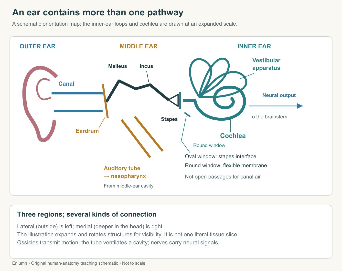
The figure expands tiny spaces so their boundaries can be seen. It does not imply that all listed structures lie in one thin section of the head. In actual anatomy, some run toward or away from the viewer. Nor should the equal visual size of two labels imply equal dimensions. Schematics trade geometrical completeness for readable relationships. Their captions should tell you which trade has been made.
Uncoil the cochlea carefully
The cochlea's spiral makes a long sensory arrangement fit within a compact region. For an introductory map, we can imagine straightening the spiral into a strip. The base is the end near the windows; the apex is the far end of the spiral. “Base” here does not mean the bottom of a picture, and “apex” does not necessarily mean its top. Rotate a drawing and those page directions change; the anatomical relationships do not.
Across a cochlear turn, three principal fluid spaces appear: scala vestibuli, scala media, and scala tympani. Scala media is the cochlear duct and contains endolymph. The other two contain perilymph and communicate near the apex through the helicotrema. The vestibular membrane separates scala media from scala vestibuli; the basilar membrane separates it from scala tympani. The sensory organ of Corti is supported on the basilar membrane. Yale Medcell, Inner Ear.
A cross-section through a coiled tube can intersect several turns. Seeing several separated circular or triangular spaces in a tissue slice therefore does not prove that the organ contains that many independent tubes. A familiar analogy is slicing through a coiled garden hose: one continuous hose may appear repeatedly in the same cut. The analogy explains the geometry only. The cochlear partitions, sensory epithelium, and ionic compartments have no counterpart in an ordinary hose.
Try a fictional paper spiral 30 centimeters long wound into three turns. A straight cut might cross it several times, depending on the cut's position. Counting crossings cannot recover the total length without additional information about the shape. Likewise, a microscope image and an anatomical reconstruction provide complementary evidence. The image shows local tissue relationships; the reconstruction helps locate those relationships within the whole organ. Neither automatically substitutes for the other.
A window need not be an open hole
The oval window and round window are interfaces at the basal region of the inner ear. The stapes footplate transmits movement at the oval window. The round-window membrane provides a compliant boundary that can move as inner-ear fluid and partitions move. Neither is an uncovered opening allowing canal air to pour into cochlear fluid. Calling them windows helps locate interfaces but can mislead if you picture two missing panes of glass.
Because fluid is difficult to compress, motion at one boundary depends on the mechanical possibilities elsewhere in the system. A sealed chamber with completely rigid walls would respond differently from one with a flexible membrane. That observation prepares the pressure-and-motion model in Chapter 2. For now, the anatomical lesson is that the boundary conditions belong on the map. A fluid space drawn without its membranes is missing part of the apparatus that determines how it moves. Purves and colleagues, The Inner Ear.
Imagine a fictional chamber with an input piston displacing two cubic units and a flexible output boundary accommodating two cubic units. If a drawing labels the input movement but omits the output boundary, it invites the reader to imagine that fluid simply vanishes or compresses without limit. The example is deliberately coarse: real cochlear motion is oscillatory and involves distributed tissue deformation. Still, it exposes why a static list of contents does not explain a functioning organ.
Locate the cell before following its message
A hair cell is a specialized epithelial sensory cell named for the bundle projecting from its apical surface. Its stereocilia are cellular projections with an actin-based internal structure; they are not strands of scalp hair. The bundle's displacement changes the cell's electrical state. A neighboring nerve ending receives synaptic input and carries action potentials onward. Receptor cell and nerve cell are therefore distinct participants, even when a small diagram compresses their connection into one arrow.
In the cochlea, the cell bodies of the primary auditory neurons lie in the spiral ganglion. The hair cells themselves belong to the organ of Corti, not to that ganglion. Supporting cells and specialized fluid-maintaining tissues are also essential. The organ cannot be understood as naked nerve endings dipped into a bowl of water. Its microscopic arrangement creates different conditions at different surfaces of a sensory cell. Purves and colleagues, Hair Cells and Mechanoelectrical Transduction.
In the bus example, the auditory pathway begins with acoustic pressure variations, proceeds through mechanical motion, and reaches cellular transduction before the resulting patterns are processed in the nervous system. At each transition, ask what is moving or changing. Air pressure is not neurotransmitter release; displacement is not an action potential; detecting a signal is not yet recognizing a familiar voice. The order matters because an explanation can fail by skipping precisely the step it was meant to clarify.
Keep the motion-sensing branch visible
The vestibular apparatus uses related sensory cells to provide information about head motion and orientation. Semicircular ducts are arranged in different planes and participate in sensing rotation. The utricle and saccule respond to forces associated with linear acceleration and gravity. Their outputs contribute to a wider system involving vision, body sensation, central processing, and motor responses. Balance is an achievement of that cooperating system rather than a liquid level in one little container. NIDCD, Balance Disorders.
Return to the person's first step onto the bus. A diagram devoted only to hearing could explain the friend's acoustic signal while saying almost nothing about stabilizing the eyes during head movement. Conversely, a vestibular map could explain an important motion input without identifying the spoken word. Both maps can include the eighth cranial nerve, the vestibulocochlear nerve, because its divisions carry information from different receptor structures. Sharing a named nerve does not make those signals interchangeable.
Consider a fictional building with an electrical cable containing separate lines for a doorbell and a temperature sensor. Damage to the outer casing does not tell you which line still works; a functioning doorbell does not establish that the temperature reading is correct. The analogy illustrates parallel pathways within shared infrastructure. It does not imply that biological signaling is a set of perfectly insulated appliance wires or that the pathways never interact centrally.
A scale check can prevent another mistake. If a fictional five-millimeter structure is drawn fifty millimeters long, the enlargement is tenfold in length. Its drawn area would be one hundred times the corresponding real area if both dimensions were enlarged equally. Neither calculation licenses comparing it with a neighboring structure drawn at a different magnification. A page combining a whole ear with a hair-cell inset therefore needs separate scale statements. The inset explains a small working arrangement; it is not evidence that a hair cell occupies a large fraction of the skull.
Read a map by testing its consequences
Suppose a student labels the external canal “filled with endolymph,” puts the ossicles inside the cochlear duct, and draws the auditory tube continuing into the brain. Three corrections are required, not a vague instruction to study harder. The canal is an air-containing skin-lined passage; ossicles belong to the middle ear; and the auditory tube reaches the nasopharynx while neural information follows nerves. Each correction restores a relationship that later physiology will rely on.
A second student's map has all those labels correct but shows light-blue arrows everywhere without explanation. It might still conceal a serious misunderstanding. Ask the student to name what each arrow represents and where its meaning changes. A reliable explanation should identify acoustic pressure, solid and fluid motion, cellular transduction, synaptic communication, and neural output. The same exercise works for the vestibular branch, beginning with head motion or orientation instead of an incoming sound wave.
Check your understanding: Why is “sound travels through the ear canal, Eustachian tube, and auditory nerve into the brain” an incorrect route?
Expected answer: The Eustachian tube branches from the middle-ear cavity toward the nasopharynx. Ordinary air-conducted hearing instead involves tympanic and ossicular motion, cochlear mechanics, hair-cell transduction and neural signaling. The auditory nerve carries action potentials, not canal air or an unchanged acoustic wave.
Application
Allow 15–20 minutes. Draw the three ear regions and use five symbols: spaces, solid structures, membranes, mechanical arrows and neural arrows. Include the external canal, tympanic membrane, three ossicles, auditory tube, two windows, cochlea, semicircular ducts, utricle, saccule and neural output.
Add a separate cross-section with the three scalae and the organ of Corti. Label the fluids and the membranes separating the cochlear duct from its neighbors. Explain why several turns in one section need not represent several separate cochleas.
Your map passes if the auditory tube does not enter the inner ear, windows are closed interfaces, the ganglion is not the hair-cell layer, and hearing and vestibular routes remain distinguishable. Use the supplied schematic and prose; do not insert objects into your ear or attempt to inspect deep structures yourself.