Hair cells and auditory information
A piano chord reaches the ear as one changing pressure waveform, not as three labeled notes traveling down separate air channels. Yet a listener may distinguish several pitches, their timing, and the identity of the instrument. The cochlea begins the analysis by responding differently along its length and converting local mechanical events into patterns of neural activity. Its operation depends on living cells that both sense movement and influence the movement being sensed. This makes the inner ear a more interesting apparatus than a passive microphone attached to a wire.
A wave has a place of greatest response
Along the cochlear partition, mechanical properties vary from base to apex. The basal region is relatively narrow and stiff; more apical regions are wider and more compliant. High-frequency stimulation generally produces its greatest response nearer the base, while lower-frequency stimulation peaks farther toward the apex. This ordered relationship is called tonotopy. It describes a pattern of sensitivity, not a set of isolated compartments each admitting only one pure tone. Purves and colleagues, The Inner Ear.
Imagine five sampling sites along a fictional uncoiled partition. A particular stimulus produces responses 1, 4, 9, 3, and 1. The third position responds most strongly, but four other positions also move. Calling the third site “the only place that hears this frequency” would discard most of the supplied observation. A different stimulus might produce 8, 5, 2, 1, and 0.5. Comparing the distributions provides more information than assigning each stimulus to one isolated point.
The diagram below shows broad response envelopes rather than a snapshot of every oscillation. An envelope describes how response magnitude varies with place. It is not the instantaneous shape of a single traveling wave, and the plotted horizontal direction is distance along the uncoiled cochlea, not the passage of time. Confusing those axes can turn a useful frequency map into an incorrect claim about the order in which a person hears the notes of a chord.
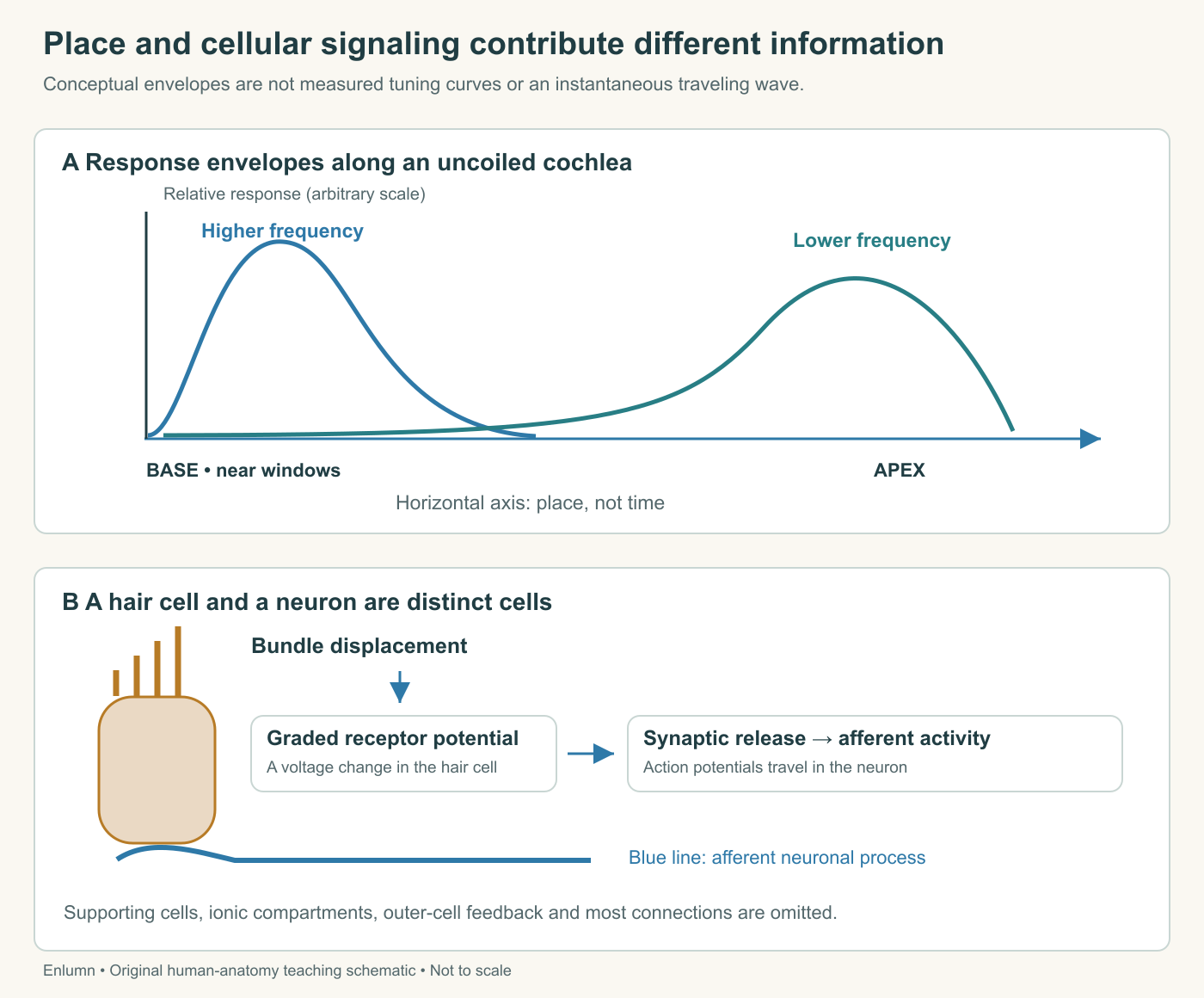
A spatial maximum is therefore only one description of the response. We can also ask how sharp the tuning is, how response grows as input increases, and how its timing relates to the stimulus. Two systems could share the same maximum position while differing on all three questions. Those differences become important when we try to explain why detecting a sound and distinguishing a sound from its neighbors are not the same achievement.
A mechanical gate changes an electrical state
The hair bundle contains stereocilia linked by fine structures that help couple displacement to mechanically gated channels. Deflection toward the tallest stereocilia increases channel opening; deflection in the opposite direction reduces it. Ionic current changes the hair cell's membrane voltage, producing a graded receptor potential. In an auditory inner hair cell, depolarization promotes calcium entry and neurotransmitter release onto nearby afferent endings. Those neurons generate the action potentials carried onward. Mature human cochlear hair cells lack the kinocilium present in vestibular bundles. Purves and colleagues, Hair Cells and Mechanoelectrical Transduction.
There are two separate transformations here. First, a displacement changes the likelihood that channels are open. Second, altered voltage changes synaptic communication with another cell. Neither step requires a strand of hair to act as a wire transmitting vibration to the brain. The term “hair cell” identifies an anatomical appearance; the explanation comes from channels, gradients, voltage, and synapses. Naming the cell is useful only if those relationships remain visible.
A fictional gate illustrates the distinction between continuous input and a changing response. Suppose 20 of 100 channels are open at one bundle position, 50 at a second, and 10 at a third. The available pathway for current changes in graded steps. Nothing in this model requires the membrane voltage to jump between only two values. Nor does 50 open channels necessarily imply exactly 50 nerve impulses; current, voltage, transmitter release, and neural firing are linked through additional dynamics.
The resting condition matters because some channels can be open before a new deflection. A signal can therefore be represented by an increase or a decrease from baseline. An observer who records only whether the response is above zero would miss that distinction. In biological signaling, baseline activity can be part of the capacity to represent change, rather than meaningless background that should always be subtracted away and forgotten.
The surrounding fluid is part of the apparatus
The bundle's apical environment differs from the environment around the cell's basolateral surface. Endolymph is potassium-rich, and the cochlear endolymphatic compartment has a positive electrical potential relative to perilymph. These conditions help drive current through open transduction channels. Specialized tissues, including the stria vascularis, maintain the environment. The arrangement depends on cellular transport, tissue barriers, and metabolic support rather than an indefinitely charged, maintenance-free battery. Yale Medcell, Inner Ear; NIDCD, How Do We Hear?.
To reason about a channel, ask both whether it is open and what drives flow through it. A doorway can be unobstructed while little passes through if there is no relevant driving difference. Conversely, a strong driving difference cannot produce the same flow through a completely closed route. The analogy is incomplete because ions respond to electrical as well as chemical differences, but it reveals why “the stereocilia moved” does not by itself specify the whole cellular response.
Suppose a teaching model gives current as conductance multiplied by driving voltage. With conductance two units and driving voltage five, current is ten units. Halving conductance at the same voltage gives five; halving the driving voltage at the original conductance also gives five. The same measured current can therefore result from two different changes. These are invented electrical units, not physiological values. The example prepares us to distinguish damaged channels from altered support conditions without diagnosing either from a single output.
Inner and outer cells have different emphases
The inner hair cells supply the dominant sensory input to the auditory nerve. Outer hair cells also transduce mechanical stimulation, but their voltage-dependent changes in length feed mechanical energy back into the cochlear partition. The protein prestin contributes to this electromotility. Active outer-cell behavior helps shape sensitivity, tuning, and nonlinear responses. It is therefore misleading to call outer cells merely spare inner cells or to describe the cochlea as entirely passive. Ashmore, Outer Hair Cells and Electromotility.
A piano-key analogy has limits here. A keyboard divides the instrument into separate keys with fixed assignments. Cochlear mechanics involve coupled tissue, distributed responses, and active feedback. A model of linked filters is closer for some questions, provided it also allows the living mechanism to change the filter behavior. No single analogy captures all of this. The right question is whether an analogy preserves the relationship being explained or quietly removes it.
Consider two supplied response curves. Model A maps input amplitudes 1, 2, and 4 to output amplitudes 1, 2, and 4. Model B maps them to 3, 4, and 5. In A, output is proportional to input. In B, each doubling of input produces a smaller proportional increase in output. B is compressive over these points. Its apparent gain, output divided by input, falls from 3 to 2 to 1.25. These invented curves illustrate nonlinearity without claiming to reproduce a person's cochlea.
This also explains why measuring behavior at one level cannot automatically predict behavior at another. If a researcher observes gain three at the lowest input and extrapolates linearly, they predict output twelve at input four. The supplied nonlinear model gives five. The disagreement is not a rounding error; it arises from a wrong assumption about the relationship. Biological measurements often require an operating range, not a single universal gain number.
An ear can return a measurable acoustic response
In 1978, David Kemp reported a human-ear acoustic response measured with signal averaging after brief acoustic impulses. A later, more slowly decaying component differed from the initial response attributed to the middle ear. The abstract reported it in the tested normal ears and not in the ears classified as having cochlear deafness, interpreting the result as evidence for a nonlinear mechanism probably within the cochlea. This was a measurement and an argued interpretation, not direct visualization of a molecular motor. Kemp, Stimulated Acoustic Emissions from Within the Human Auditory System.
The methodological lesson deserves attention. A microphone near the canal records pressure, not hair-cell length or nerve impulses. To infer an internal source, investigators must consider the timing, stimulus behavior, equipment response, and alternative mechanical explanations. Repeated measurements and averaging can help reveal a small consistent response, but they do not automatically remove a systematic artifact. A recurring artifact can survive averaging just as a recurring biological signal can.
Here is a small averaging example to make the measurement problem tangible. At one fixed delay after a stimulus, suppose four invented recordings read 2, 6, 3, and 5 units. Their mean is four. If a stable response contributes four units and the remaining departures are random, averaging helps estimate that recurring contribution. But if the equipment itself contributes two units on every repetition, averaging preserves those two units as well. A control recording is needed to investigate that possibility.
Timing alignment is another assumption. If the biological response shifts between repetitions, averaging at a fixed delay can blur its shape. The resulting small average does not necessarily mean that every individual response was small. Researchers therefore need to distinguish weak responses from inconsistent timing and from instrument noise. These invented numbers do not reconstruct Kemp’s data; they explain why a method capable of revealing a response still needs an account of what its measurement means.
The inspected abstract does not provide a participant count, so we should not invent one. Nor does “present in all tested normal ears” establish that every healthy ear in every condition must produce a detectable response. The study's observation belongs to its particular protocol and sample. Modern interpretations draw on additional evidence, including outer-cell mechanics, but a historical paper should not be retroactively credited with directly measuring everything later work helped establish.
Timing adds information that a place map cannot contain
Auditory nerve activity can preserve information about stimulus timing through patterns of firing relative to a waveform. Phase locking means spikes tend to occur around particular phases; it does not require a fiber to fire on every cycle. Place-related information and timing-related information can coexist. There is no need to imagine a switch that turns off all place coding whenever temporal information is available. Purves and colleagues, Tuning and Timing in the Auditory Nerve.
For an original example, mark repeated cycles at times 0, 2, 4, 6, and 8 milliseconds. A hypothetical fiber fires near 0, 4, and 8, skipping intervening cycles. Another fires near 2 and 6. Neither fires every cycle, yet both show a relationship to the same two-millisecond rhythm. Counting each fiber's average spikes alone loses that temporal structure. The example is a diagram exercise, not a claim that a particular human nerve follows exactly this schedule.
Timing patterns also matter within a complex sound. Two invented sequences can contain the same total number of events but in different orders: short–short–long versus long–short–short. A total count treats them as equal; an ordered record does not. In speech or music, changing temporal relationships can change what is recognizable even when broad energy measures remain similar. To explain auditory information, we need descriptions that retain the distinctions the task depends on.
The cochlea prepares information; the nervous system keeps working
Primary auditory neurons reach cochlear nuclei in the brainstem. Subsequent auditory pathways include bilateral processing and structures such as the inferior colliculus and medial geniculate nucleus before cortical processing. Tonotopic organization continues, but it does not imply that one cortical cell hears a whole song. Information from the two ears, prior experience, attention, and task demands contribute to what a person can distinguish and recognize. Purves and colleagues, Auditory System Summary.
A microphone can detect a pressure pattern without knowing whether the speaker is a friend. Likewise, successful receptor transduction does not alone explain recognition. A complete account must connect physical availability to neural representation and then to the task being performed. The distinction protects us from two mistakes: treating perception as if it requires no functioning peripheral apparatus, and treating a functioning peripheral apparatus as if it contains the whole explanation of understanding.
Check your understanding: A hypothetical fiber fires on every second cycle at approximately the same phase. Is it phase locked, and does its average firing rate alone preserve the original cycle period?
Expected answer: It can be phase locked despite skipping cycles. The timing relative to the stimulus carries information that an average firing rate alone does not uniquely preserve; the same average could arise from other temporal patterns.
Application
Allow 20 minutes. Draw an uncoiled cochlear map with base, apex, and two broad response envelopes. State explicitly that the horizontal axis is place, not time. Then draw bundle displacement → graded receptor potential → synaptic release → action potentials in an afferent neuron.
Use the supplied nonlinear curve to calculate gains at inputs 1, 2, and 4: 3, 2, and 1.25. Explain why extrapolating the first gain to the largest input fails. Finally, write 150 words distinguishing what Kemp's microphone measured from the internal mechanism he inferred. A successful answer does not invent a sample size, equate hair-cell voltage with a nerve impulse, or claim one cell hears a complete complex sound.