Focusing and Controlling Incoming Light
A lamp can make a blurred page brighter without making its letters sharp. Turning a focusing control can sharpen an image without admitting more light. These familiar optical distinctions are easy to lose when all of the eye's adjustments are described as “helping us see.” In this chapter, we separate where rays converge, how many are admitted, and how the living eye changes its focusing arrangement.
The retina lies at a particular location inside the globe. Light arriving from different object distances must be brought into useful focus near that surface. The eye cannot solve every change by moving the retina back and forth like a screen on a laboratory rail. It changes optical power, principally through the lens, while controlling the pupil and coordinating where the two eyes point.
What a ray diagram represents
A ray is a line used to represent the direction of light propagation in an optical model. It is not a nerve, a stream of visible pigment, or the outline of an object. Light from one point on a page can reach different parts of the pupil. Focusing brings those admitted rays together near a corresponding image point. Repeat this relationship across many object points and a spatial image is formed.
Start with an original paper model: a point at the top of a vertical arrow, a converging optical element, and a screen beyond it. Draw one ray through the center of an ideal thin lens and another initially parallel to its axis. After the lens, direct the second ray through the far focal point. Their intersection locates the image of the arrow's top within this approximation.
The resulting real image is inverted. That does not mean a small observer inside the brain sees an upside-down picture and rotates it. Neural activity preserves and transforms spatial relationships; perception develops from the processing of that activity. Image orientation is an optical relationship, whereas knowing which way is up depends on a wider biological system.
A useful diagram must show more than two crossing lines. Label the object, optical element, image plane, and direction of propagation. State whether you are illustrating a distant object or a near one. A pair of lines that meet somewhere behind a drawn lens does not establish that they meet on the retina.
Refraction depends on an interface
Refraction is the change in direction that light can undergo when crossing between media with different refractive indices. Refractive index describes how light propagation in a material compares with propagation in a vacuum. At normal incidence, a ray can pass through an interface without changing direction; at other angles, the index difference and surface orientation determine bending.
The corneal system contributes much of the eye's optical power. The lens contributes additional, adjustable power. Purves and colleagues' account of image formation explains the large corneal contribution and the lens's role in accommodation. A transparent structure does not automatically have strong focusing power: curvature and the refractive-index relationships across its surfaces matter.
Compare two original models with exactly the same curved transparent shape. Place one in air and the other in a surrounding medium whose refractive index nearly matches its own. The shape is unchanged, but the index contrast is smaller in the second model, so its refracting effect changes. You cannot infer optical power from a photograph of shape alone without knowing the surrounding media.
This is why the common phrase “the lens focuses all the light” is incomplete for the human eye. It overlooks the cornea and the interfaces encountered before the lens. Yet replacing it with “the cornea does everything” would also fail: the smaller adjustable contribution can be decisive when object distance changes. Dominance in a total and importance in an adjustment are different questions.
Use a simple equation without mistaking it for an eye
For an ideal thin lens in air, the relationship between focal length, object distance, and image distance can be written as 1/f = 1/u + 1/v. Here f is focal length, u is object distance, and v is image distance, using positive values for the real object and real image in the examples below. This equation is a simplified optical model. A human eye contains separated refracting surfaces and different internal media.
Suppose the model lens has a focal length of 50 millimeters and the object is 200 millimeters away. Then 1/v = 1/50 − 1/200 = 3/200, giving v approximately 66.7 millimeters. A screen placed at 50 millimeters would not intercept the sharp image of that near object. Keeping the screen fixed requires changing the optical arrangement.
Now put the object at 100 millimeters while leaving focal length unchanged. The equation gives v = 100 millimeters. Moving the object closer has shifted the image farther behind the lens. This does not mean that near objects physically move a person's retina backward. It identifies the focusing demand that accommodation must address in a biological system with a relatively fixed retinal surface.
Finally, keep the screen at 60 millimeters and place the object at 300 millimeters. The needed focal length is 50 millimeters because 1/300 + 1/60 = 1/50. Move the object to 150 millimeters while keeping the screen at 60. The required focal length becomes approximately 42.9 millimeters. The model needs greater converging power for the nearer object.
Optical power in diopters is the reciprocal of focal length in meters for this simple lens convention. A 0.050-meter focal length corresponds to 20 diopters. A 0.0429-meter focal length is approximately 23.3 diopters. The difference is about 3.3 diopters, but these are model lens values, not a prescription or the complete power of a real eye.
Unit discipline prevents striking errors. Taking the reciprocal of 50 millimeters and reporting 0.02 diopters would mix units. Convert to meters first. More generally, attaching a familiar medical unit to an invented model does not turn its result into individualized medical information.
Accommodation is a mechanical relationship
In the conventional introductory account, ciliary-muscle contraction reduces tension in the zonular arrangement, allowing the lens to become more rounded for near focus. Relative relaxation permits greater flattening through zonular tension. The lens changes shape; the zonules transmit mechanical forces. Neither structure is a tiny motorized glass lens sliding along a rail.
The apparently surprising step is that muscle contraction can reduce tension in another structure. Imagine a ring whose inward movement brings the attachment points of radial supports closer to the central object. The supports can become less taut even though the ring's muscle is contracting. The direction and geometry of force transmission matter more than the word “contraction” alone.
The actual ciliary and zonular apparatus is more complex than that ring model. Its value is to preserve the central causal sequence without pretending that every fiber experiences identical forces. A sound introductory explanation specifies muscle activity, changed tension, changed lens shape, and changed optical power as separate links.
Read the mechanism arrows independently from the light rays. A mechanical arrow does not show a photon's path. This practice prevents a familiar diagram error in which light appears to travel through a muscle before reaching the lens.
Effort, movement, and optical response are different measurements
In a human MRI study, Strenk and colleagues studied 25 adults aged 22–83 under two accommodative stimulus conditions. They measured dimensions of the ciliary-muscle ring and lens. Muscle contraction remained present in the participants, including older adults. The observation argues against explaining age-related loss of near focusing simply as complete disappearance of ciliary-muscle activity.
The study's stimulus demanding substantial accommodation was not proof that every participant achieved that optical response. A target establishes a demand; imaging establishes selected anatomical changes. The abstract's preferred theory of presbyopia was an interpretation of findings, not a final settlement of every mechanical contribution. The bounded lesson is to measure the different links rather than substitute effort for outcome.
Construct a fictional chain with four boxes: near target, neural command, muscle movement, optical focus. Suppose a supplied observation confirms movement in the third box while focus remains inadequate in the fourth. The observation rules out “no movement at all” in that case, but it does not identify every possible problem in force transmission or lens response.
This reasoning applies throughout physiology. An active pump may deliver little flow against an obstruction; an electrical command may produce a limited mechanical response. In the eye, asking whether a person tried to focus, whether a muscle moved, and whether an image sharpened produces three different kinds of evidence.
A pupil is an adjustable aperture
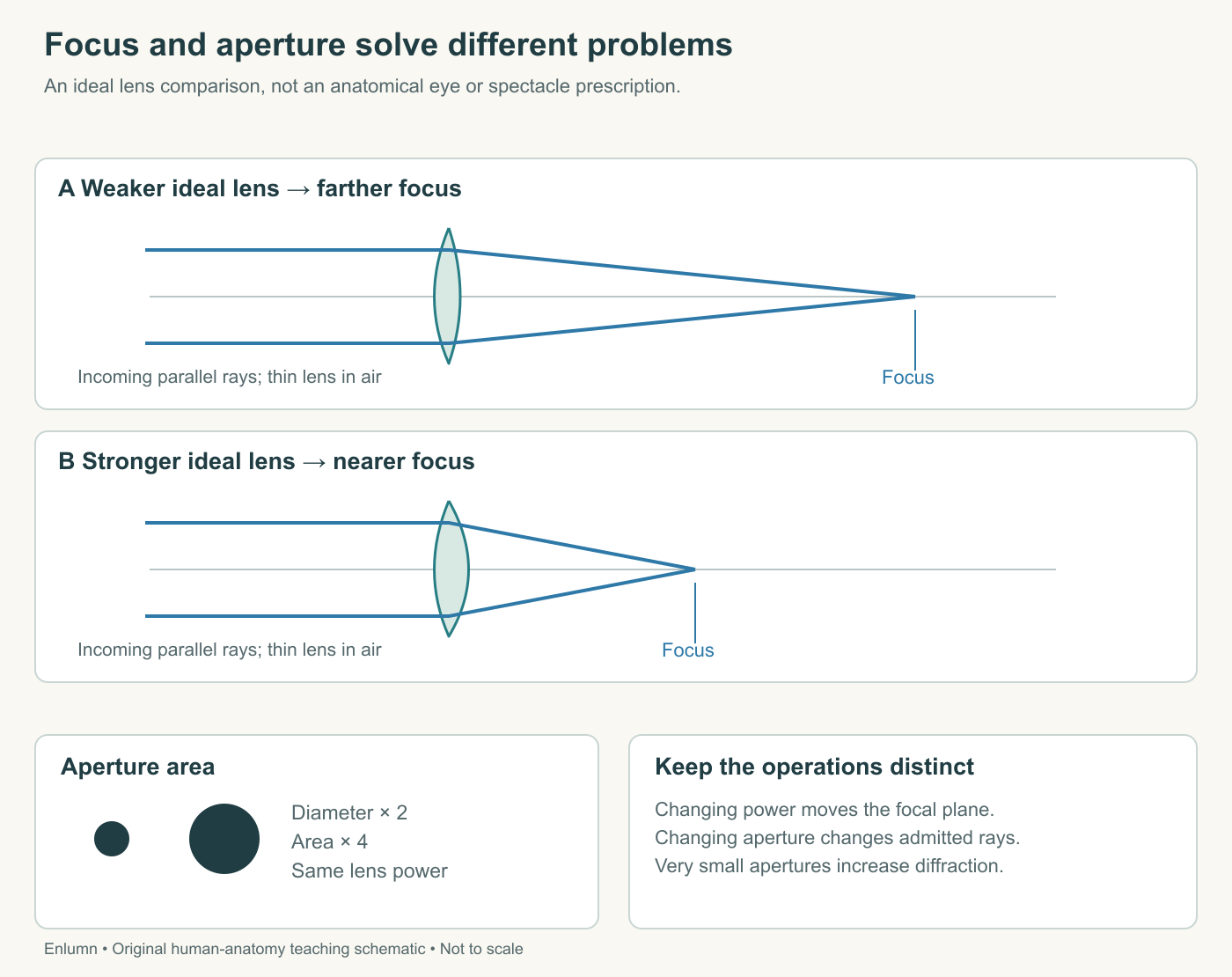
The iris's muscles alter pupil size under neural control. Changing the diameter changes the opening's area. For a circular aperture, area is proportional to diameter squared. A fictional change from a 2-millimeter diameter to 4 millimeters produces four times the opening area, not twice. Under specified constant illumination and transmission conditions, that allows a larger quantity of light into the model system.
Changing from 3 to 6 millimeters gives the same fourfold area ratio. Changing from 3 to 4 gives a ratio of 16/9, about 1.78. These calculations isolate geometry. They do not establish identical retinal illumination in every eye, because optical transmission and other conditions can differ.
An aperture also restricts which ray paths enter. A smaller opening can reduce the size of a defocused blur patch and increase the range of object distances that appear acceptably sharp. This range is called depth of field. It does not mean the lens has acquired every focal length at once. Rather, the criterion for acceptable blur is met over a broader range.
Draw a cone of rays meeting behind a screen. The screen intercepts a patch rather than a point. Narrow the admitted cone while keeping its meeting point fixed. The patch at the screen becomes smaller. You have improved the blur geometry without moving the true focus onto the screen. This is the distinction between restricting rays and changing optical power.
Smaller is not always better
A small aperture admits less light. At sufficiently small openings, diffraction—the spreading associated with light's wave behavior—also limits resolution. Larger openings admit more light but can increase the influence of optical aberrations, departures from ideal image formation. The outcome is a tradeoff, not a rule that the smallest possible pupil always produces the best vision.
The term “sharp” itself needs a criterion. An observer might judge whether a letter is readable, whether two points can be distinguished, or whether faint contrast is visible. Different tests can respond differently to changes in illumination, aperture, focus, and neural processing. One measure cannot stand in for every aspect of visual performance.
For a supplied example, model A admits 100 light units and produces a blur patch two units wide. Model B admits 25 light units and produces a patch one unit wide. If the task is resolving a bright edge, B may offer useful spatial improvement. If the task is detecting a very faint signal, its reduced light could be costly. Without specifying the task and noise, “which image is better?” has no complete answer.
None of these comparisons calls for looking at a bright lamp, staring into the sun, or manipulating the eye. The geometry can be understood from supplied drawings. A physical exposure is unnecessary to establish a relationship between diameter, area, and a ray cone.
Name refractive differences precisely
Myopia describes an optical situation in which distant-object light focuses in front of the retina when accommodation is relaxed. Hyperopia places that focus behind the retina under the corresponding condition. Astigmatism involves differences in optical power across orientations, so one simple focal-point model is insufficient. Presbyopia is the age-related decline in accommodative ability, affecting near focusing.
The NEI's refractive-error overview distinguishes these categories. Hyperopia does not guarantee effortless clear distance vision, and presbyopia is not simply another name for a short globe. More than one condition can coexist. These definitions identify optical relationships; they do not diagnose the cause of an individual's blur.
A corrective lens alters rays before they reach the eye. It can improve the match between the combined optical system and retinal location without rebuilding retinal neurons. That is why correcting focus and restoring a damaged neural pathway are different tasks. The final chapter will compare those possibilities using supplied evidence.
Check your understanding: A smaller aperture makes a fictional blurred image more readable. Does this prove that its lens changed optical power? What happens to opening area when diameter is halved?
Expected answer: No. Restricting admitted rays can reduce the blur patch and increase depth of field without changing the true focal position. Halving diameter reduces circular opening area to one quarter, under the same geometrical assumptions.
Application
Allow twenty minutes for a paper exercise. Use an ideal thin lens in air with a screen fixed 80 millimeters behind it. Calculate the focal length needed for an object 240 millimeters away, then for one 160 millimeters away. Convert each focal length to optical power in diopters. Do not treat the results as spectacle prescriptions.
Draw a separate pair of ray cones showing how reducing aperture can shrink a blur patch at an unchanged screen and focal point. Label the light lost as well as the smaller blur. Finally, write four sentences distinguishing a near target, muscle contraction, lens-shape change and achieved focus.
The model focal lengths are 60 and approximately 53.3 millimeters, corresponding to about 16.7 and 18.75 diopters. A strong explanation preserves the distinction between focus and aperture, notes that light quantity also changes, and treats the human MRI findings as measurements of selected structures rather than proof of a complete optical response.