A microscopic working arrangement
Draw a hexagon, place a vein in its center, and put three small circles at each corner. You have the beginning of a familiar liver diagram. Now open a human liver slide. The neat outline is difficult to find, channels meet at uneven angles, and a tissue slice shows only part of each branching structure. The diagram has not become useless. Its purpose is to make a relationship intelligible, and the real tissue asks us to distinguish that relationship from the geometry we used to teach it.
A lobule is a way of organizing a view
The classic hepatic lobule groups tissue around a central venous outflow. Portal regions near its periphery contain branches of the portal vein and hepatic artery together with bile ducts. Blood passes from these incoming regions through sinusoids toward central drainage. Hepatocyte plates occupy the spaces between the vascular channels. This is the organizing relationship behind the hexagon.
A human liver is not a box packed with identical six-sided tiles. Lobular boundaries are often much less conspicuous than the teaching outline, and a slice can intersect a structure at an angle or miss a supposed corner. Adjacent regions share branching networks rather than functioning as isolated miniature livers. The outline helps trace flow; it does not prove that every cell belongs to a neatly walled compartment.
The term portal triad highlights three structures: a portal venous branch, a hepatic arterial branch and a bile-duct profile. An actual portal tract includes connective tissue and may contain lymphatic vessels and nerves, as well as multiple profiles of the highlighted structures. “Triad” is a reminder of three kinds of route, not a promise that every microscope field contains exactly three circular openings.
This matters when identifying a central vein. A large lumen alone is insufficient. A venous branch within a portal tract can also have a large lumen. Its neighbors help distinguish it from central venous drainage: a portal tract has associated duct and arterial profiles, whereas a central vein receives sinusoids from surrounding tissue. Anatomy becomes more reliable when relationships support a label rather than size doing all the work.
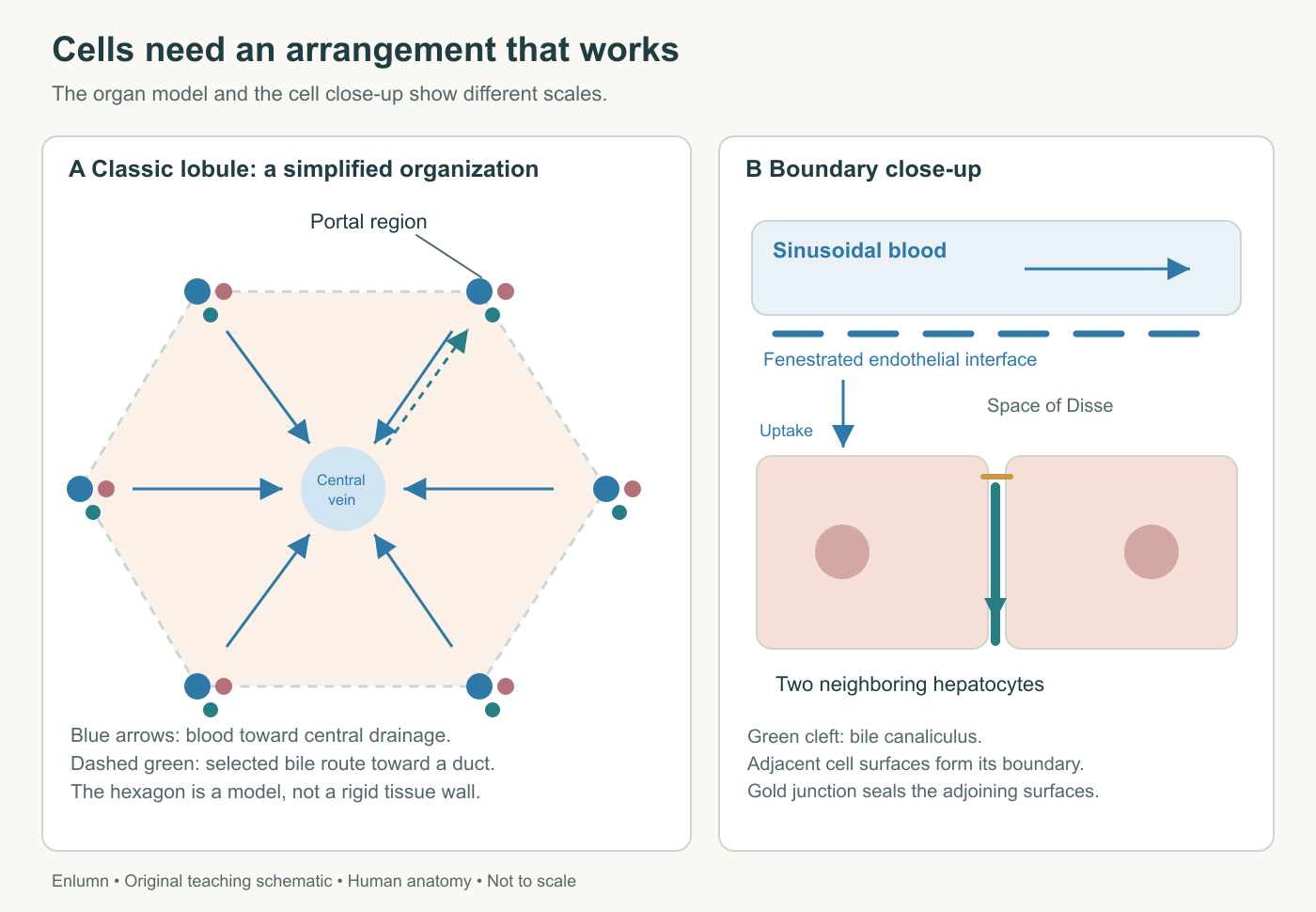
The classic lobule is a drainage model, not a rigid hexagonal wall. The close-up separates blood, the endothelial interface, the space of Disse and hepatocytes. The canaliculus is a sealed secretory cleft; its downstream route continues beyond this small section. Blood and bile do not share one open space.
Look at the human section
Open the Histology Guide human liver slide, MH 126b. Select Hepatocytes in its description and allow the detailed image to load. Large pink cells with conspicuous purple nuclei are arranged in branching plates. Paler channels separate them. Some contain red blood-cell profiles; elongated dark nuclei occur along parts of the channel boundaries. The atlas notes prominent lipofuscin in this specimen, visible as brownish granules in many cells.
Then select Portal Triads. Compare the cluster of profiles in connective tissue with the surrounding hepatocyte plates. Duct profiles have a recognizable epithelial lining, with nuclei arranged around or along a lumen; vascular profiles have different wall organization. An oblique section can make a lumen appear elongated, small or absent from the visible plane. Do not make perfect circularity part of your definition of a duct.
The stain supplies contrast, not natural color. It also does not directly show transport across membranes. You can identify visible cell outlines, nuclei, channels and spatial relationships, but you cannot watch bile being secreted in this fixed section. Very small structures, including the detailed barrier between a canaliculus and surrounding tissue, require other preparations or methods. The microscopic view and the mechanism diagram answer different questions.
A useful sketch records three things separately: what is visible, what the atlas identifies, and what physiology leads you to infer. For example, the visible pale channel supports a structural observation; the identification of that channel as part of the sinusoidal system uses its context; the direction of flowing blood is physiological knowledge, not motion recorded in the still image. Keeping these levels distinct makes your interpretation stronger, not less decisive.
One hepatocyte, different surfaces
A hepatocyte is a polarized epithelial cell. Polarity means that different regions of its surface have different arrangements and jobs. Portions face the blood-associated extracellular environment. Other portions form the boundaries of bile canaliculi with neighboring hepatocytes. Transport to blood and transport to bile therefore occur at different membrane domains within the same cell.
Imagine two adjoining hepatocytes in a cross-section. A small canalicular space lies between selected parts of their adjacent surfaces. Junctions help separate that space from other extracellular compartments. Bile enters the space from the cells and travels onward through the network. The canaliculus is not an independent pipe with a complete ring of separate duct cells at this stage; its boundary is made by the hepatocytes themselves. Larger downstream ducts have their own epithelial lining.
This arrangement explains how one cell can take up a substance from the blood side, change it internally and secrete a product toward bile. The path crosses the cell rather than simply continuing through an open hole from sinusoid to canaliculus. A transporter at one membrane is not interchangeable with a transporter at another. Its position helps determine the direction in which a molecule can be handled.
For an original thought experiment, suppose a cell makes a product normally but the mechanism that exports it at its canalicular surface fails. The production pathway could remain active while outward delivery falls. Conversely, an export mechanism could work normally but have little to transport because synthesis has fallen. Measuring the amount outside the cell alone does not distinguish these two failures. A mechanistic explanation needs both the internal process and the boundary step.
A further complication is that a cell's surface is three-dimensional. A two-dimensional textbook drawing may show one blood-facing side and one bile-facing side, like the front and back of a house. Actual hepatocytes can contact several neighbors and vascular spaces. The simplified division remains valuable, but rotating or slicing the cell changes which membrane regions are visible. A flat illustration should preserve polarity without pretending to exhaust its geometry.
Exchange across the sinusoidal interface
The blood in a sinusoid is separated from hepatocytes by a specialized endothelial interface. Endothelial cells line vascular spaces. In the liver, their fenestrations and the organization of the surrounding tissue facilitate exchange of plasma constituents. Fenestrations are small openings in these cells; they do not mean that a red blood cell normally exits the vessel to enter a hepatocyte.
Between the endothelium and the hepatocyte surface is the space of Disse, a narrow extracellular region involved in exchange. Hepatocyte microvilli extend toward this space and increase the available surface. Plasma constituents can approach that surface, while actual entry into a hepatocyte still depends on their properties and the relevant membrane pathways. Accessibility and uptake are successive questions.
Lautt's microcirculatory account describes this relationship between sinusoidal blood, the perisinusoidal space and hepatocyte contact. It gives a reason for thinking beyond bulk blood flow: even a well-supplied organ needs an effective interface between circulating material and its cells. A change in that interface can matter without eliminating the whole vessel.
Here is a simple geometric model. Imagine a fixed tissue volume divided into many thin plates rather than one thick block. More of the tissue can lie near an exchange surface. That reduces the distance over which supplied molecules must travel to reach cells, provided blood actually reaches the intervening channels. The model does not claim that surface area alone determines function. Flow, concentration, permeability, cellular uptake and metabolic demand all remain relevant.
Separate those influences with an invented comparison. Region A has ample flow but poor passage across its interface. Region B has a permeable interface but very little flow. Both can have low delivery to cells. Opening more membrane transporters in B would not manufacture the missing supply; increasing flow in A would not necessarily correct an ineffective barrier. The same low uptake measurement can arise through different upstream causes.
The other cells are active participants
Kupffer cells are resident macrophages associated with the sinusoidal blood compartment. They participate in handling particles, cellular remnants and microbial material, and in immune signaling. They are not a second name for hepatocytes. Their position gives them access to material reaching the liver through the circulation, but they do not simply sterilize everything before it returns to the body.
Hepatic stellate cells occupy a perisinusoidal position and have roles in matrix maintenance and vitamin A storage in their resting state. During injury they can change their behavior and contribute to collagen-rich scar formation. The physiological review by Puche, Saiman and Friedman describes this change toward a contractile, matrix-producing state. We will use that relationship in the final chapter without treating the transition as a single switch shared identically by every cell.
Cholangiocytes line the bile ducts. They modify the fluid passing through them, including through regulated movement of ions and water. A duct is thus more than a passive drain. A hepatocyte may form the initial secretion, while the downstream epithelium changes its composition and volume. The bile-secretion reference distinguishes these cellular contributions.
Finally, the extracellular matrix is the material outside cells that helps organize and support tissue. It includes proteins with mechanical and signaling roles. Normal matrix is not synonymous with pathological scar. A healthy organ requires organized support; disease can alter its quantity, composition and distribution. “Less matrix is always better” would be as mistaken as “more support always improves function.” The important question is whether the arrangement supports the necessary exchanges and routes.
Location changes the conditions a cell experiences
Blood does not have the same composition at every point along a sinusoidal path. Cells remove some substances and add others. Oxygen availability generally declines as blood moves away from incoming regions toward venous outflow. Local signals and patterns of gene activity also differ. This spatial variation in metabolic activity is called zonation.
The acinus is another way to organize the tissue conceptually, emphasizing the area supplied by a small incoming vascular branch. Its commonly numbered zones run from the incoming periportal region toward the later-perfused pericentral region. The classic lobule is centered on a venous exit; the acinar view emphasizes supply. The word “center” can therefore refer to different landmarks in different models. Read the legend before deciding which way blood flows.
It would be an error to interpret zonation as three tiny organs performing mutually exclusive jobs. Patterns overlap, can change with conditions, and involve gradients rather than perfectly sharp lines. Nor does lower downstream oxygen mean that the last cells normally receive none. It means their circumstances differ from those of earlier cells and can make certain disturbances affect locations differently.
Use a fictional two-region calculation to see why an average can conceal this. Suppose equal-sized regions A and B contain 8 and 2 units of a stored material per tissue unit. The average is 5. Another liver model with 5 in each region has the same average but a different distribution. A bulk measurement cannot distinguish them. If an enzyme or supply limitation differs by region, the two models could respond differently despite starting with an identical whole-organ average.
Now let a channel deliver 100 units of a tracer and let the first region remove 20. The second region receives 80, not the original 100. If it removes a quarter of what reaches it, that is another 20, leaving 60. Its extraction fraction is larger than the first region's 20%, but the absolute amount removed is the same. This arithmetic separates location, fraction and amount. It is not a measured oxygen profile or a claim that a real sinusoid has two discrete chambers.
Architecture is part of function
The chapter's different views now fit together. Thin hepatocyte plates place working cells near a specialized vascular interface. Polarized surfaces separate blood-associated exchange from biliary secretion. Portal tracts and central drainage give the network direction. Other cell types maintain boundaries, handle material, modify bile and reshape the matrix. None of these features alone performs “liver function.” Their relationships make multiple functions possible.
Return to the original hexagon. It can still be useful if it tells you where inflow, outflow and bile drainage belong. Add a microscopic inset to show the endothelial interface, space of Disse and canaliculus. Then place the human slide beside it and identify what the drawing simplifies. A model earns its place by helping you explain the tissue; the tissue should not be forced to match the model's neatness.
Check your understanding: Why does an intact bile duct not prove that bile secretion from hepatocytes is normal?
Expected answer: The duct is a downstream route. Initial bile formation also requires polarized hepatocyte membranes, appropriate transport and an intact canalicular network. A patent duct cannot establish that these upstream steps work. Conversely, normal cellular synthesis does not establish successful export or downstream flow.
Application
Allow 12–15 minutes. Draw a classic lobule and a magnified pair of hepatocytes. Label a portal tract, central vein, sinusoid, endothelial interface, space of Disse and bile canaliculus. Use different arrows for blood flow, uptake into a hepatocyte and bile secretion.
Inspect the linked human slide's Hepatocytes and Portal Triads views. Record three visible features and two functions that the still image cannot establish. Finally, use the chapter's two-region tracer calculation to explain why a fraction removed and an amount removed are not the same measurement. Keep this diagram for the final course task.