Signaling the state of fuel supply
Our meal is supplying absorbable products to the intestine, but the body's needs do not wait for digestion to finish. Some tissues are using glucose, others are storing or releasing material, and the liver is changing several pathways at once. The endocrine pancreas participates in coordinating these flows. Understanding that coordination requires more than remembering that insulin lowers glucose and glucagon raises it: we need to identify the cells, signals, targets and returning effects.
A concentration is the result of competing rates
Blood glucose concentration describes glucose relative to a volume of blood or plasma. Its change reflects the balance between entry, removal and distribution. Intestinal absorption supplies glucose after a carbohydrate-containing meal; the liver and kidneys can contribute glucose from endogenous processes. Tissues remove glucose for use or storage. A steady concentration can therefore coexist with substantial movement through the system.
Imagine an invented pool containing 100 glucose units in a fixed volume. During one interval, 12 units arrive from intestinal absorption and 8 from endogenous release, while tissues remove 20. The pool remains at 100. If endogenous release falls to 3 while absorption stays at 12 and removal stays at 20, the pool falls to 95. That change did not require the intestine to stop absorbing or the pancreas to remove glucose directly from blood.
Real glucose regulation is not a single well-mixed bucket, and the rates themselves respond to conditions. Nevertheless, the bookkeeping prevents a common error: assigning every change in blood glucose to food entering the body. Endogenous production, tissue uptake, distribution and, in some circumstances, urinary loss also matter. A hormone can affect the measured concentration by changing more than one side of the balance.
Beta cells connect metabolism to secretion
An insulin-producing beta cell responds to glucose partly by metabolizing it. Glucose crosses its membrane through transport proteins and is phosphorylated by glucokinase. Subsequent metabolism alters the cell's energy state, including the balance of ATP and ADP. This couples the availability of a nutrient to a change in cellular signaling rather than requiring a tiny glucose meter inside the cell.
A central triggering mechanism involves ATP-sensitive potassium channels, often written KATP channels. Under conditions favoring their closure, less outward potassium current passes through them. The membrane can become less negative inside relative to outside: it depolarizes. Voltage-dependent calcium channels then open, allowing calcium to enter. Increased calcium near release machinery helps trigger fusion of insulin-containing granules with the cell membrane.
The Endotext insulin account explains this link among metabolism, electrical activity and secretion. It is a selected pathway, not the entire regulation of a beta cell. Other nutrients, hormones and neural signals modify the response, and granule availability and cellular condition also matter. Closing a channel is one event within a working cell, not an independent guarantee of a specified quantity of insulin.
Human glucose transport deserves care. Direct work on human beta-cell preparations identified a major role for GLUT1, while glucokinase contributes to glucose sensing. Introductory diagrams often label a generic beta cell with GLUT2 alone. We will leave the transport step broadly labeled in our main mechanism diagram rather than turn an oversimplified transporter label into the explanation of human secretion. The selected human finding supports that choice; no comparative animal lesson is required.
Follow the direction of each event. Glucose enters and is metabolized; a potassium conductance decreases; the membrane depolarizes; calcium enters; granules release their contents. Insulin is not made from incoming glucose during those few steps. It is a protein synthesized from amino acids through gene expression and processing. Stimulated release can draw on previously prepared granules, while synthesis and replenishment have their own time course.
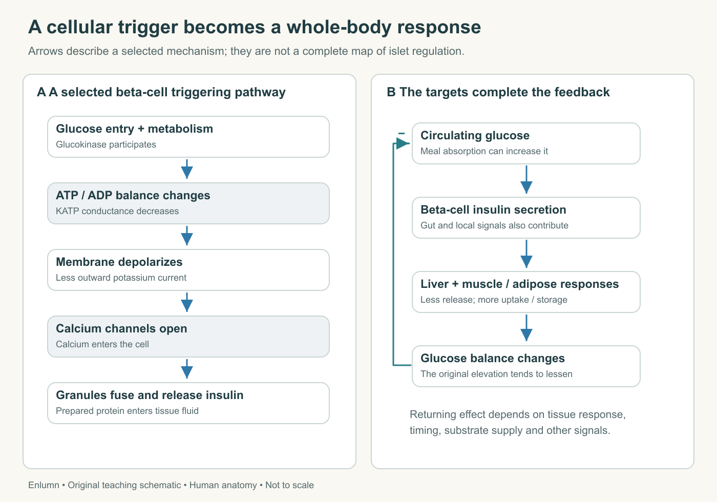
Read the left panel as a sequence of different kinds of event: nutrient metabolism, altered conductance, membrane depolarization, calcium entry and granule fusion. The right panel changes scale to a whole-body relationship. The returning minus sign means that the response tends to oppose the original elevation; it does not mean glucose becomes negative. The target box combines several tissue-specific actions, so unpack it into hepatic release and muscle/adipose uptake when explaining the mechanism. Alpha cells, other hormones and detailed neural influences are omitted from this selected insulin loop.
Manufacturing the signal is a separate task
Insulin is initially produced as a larger precursor. Processing and folding through the secretory pathway yield mature insulin and C-peptide, a connecting segment removed from proinsulin. The two products are released together in approximately equimolar amounts during normal processing. Equimolar means equal numbers of molecules, not equal masses: differently sized molecules can be present in the same count while weighing different amounts.
That distinction creates a useful measurement opportunity, developed further in Chapter 4. It also clarifies why granule release and new protein synthesis must not be drawn as one arrow. If a beta cell has a stock of mature granules, its initial release can increase before an equivalent amount of new insulin has been manufactured. Continued secretion, however, depends on maintained production, processing and cellular health.
Use an invented granule store of 80 release-ready units. During an interval, 20 units are released and 5 newly prepared units enter the store. It ends at 65. If only the final stock is measured, the 15-unit decline does not show that secretion was 15: replenishment occurred at the same time. If preparation rises to 20, the stock can remain unchanged while secretion continues at 20. A stable store is compatible with substantial work.
This is the same conservation principle used in other courses, applied to a new biological problem. The point is not that every organ behaves like the same bucket. Here the extra question concerns readiness: total stored protein and granules immediately available for release are not necessarily the same pool. A mechanism can fail through impaired synthesis, processing, transport or release even if some insulin-related material is still detectable.
Insulin changes what target tissues do
Insulin binds receptors on responsive cells and activates intracellular signaling. In skeletal muscle and adipose tissue, one important response recruits GLUT4 glucose transporters to the cell surface, increasing the capacity for glucose entry under suitable conditions. The transporter and the insulin receptor are different proteins. Insulin does not carry a glucose molecule through the membrane like a delivery vehicle.
The liver responds differently. Insulin helps favor glucose use and storage and suppresses hepatic glucose production through coordinated direct and indirect effects. Hepatic glucose transport is not explained by the same GLUT4 recruitment mechanism used for muscle. The cell biology of systemic insulin function distinguishes pancreatic secretion, vascular delivery and tissue responses. A useful account preserves those tissue-specific mechanisms instead of declaring that insulin opens every cell in the body.
Many cells take up glucose without requiring an acute insulin-triggered GLUT4 response. Even in insulin-responsive muscle, contraction and other conditions can alter uptake. Therefore, reduced insulin action does not mean no glucose enters any cell anywhere. Nor does adequate entry guarantee normal downstream metabolism. “Insulin helps regulate glucose handling” is accurate precisely because it leaves room to specify which tissue and which process changed.
Insulin also influences protein and lipid metabolism. During nutrient availability, storage and synthetic pathways often become more prominent, while mobilization of stored fuel is restrained in relevant tissues. These are coordinated changes in rates, not a declaration that every molecule must now be stored. A person walking after a meal can continue using fuel while other tissues adjust storage. Physiological states overlap rather than following a single whole-body on–off command.
Glucagon supports a different part of the balance
Alpha cells release glucagon, a peptide hormone with important actions on the liver. It promotes hepatic glucose output, including glycogen breakdown and support for gluconeogenesis. The relative contribution depends on available stores, precursor supply, timing and other signals. A hormone cannot release glycogen that is no longer present or produce glucose without usable substrates and energy.
The Endotext glucagon review describes its hepatic actions and regulation by glucose, amino acids and local signals. Low glucose can stimulate glucagon release, but glucagon is not solely a fasting hormone. Amino acids from a protein-containing meal can also stimulate it. Our rice-and-bean meal supplies several kinds of nutrient, so predicting its entire response from carbohydrate alone would discard relevant information.
Insulin and glucagon can both increase in some meal contexts. Their actions help coordinate amino-acid handling, glucose availability and other processes. Calling them simple opponents is useful for one part of glucose regulation but becomes misleading if extended to every tissue and nutrient. Glucagon's principal glucose-mobilizing target in this introductory account is the liver; do not draw it directly releasing glucose from skeletal-muscle glycogen into blood.
The alpha cell also receives influences from neighboring islet cells and neural pathways. Delta-cell somatostatin can restrain local secretion, and beta-cell products participate in the local environment. These paracrine effects occur among nearby cells. They help explain why an intact islet's behavior cannot be inferred entirely from an isolated cell given one nutrient. Local communication, perfusion and the wider circulation all contribute.
Close the loop without inventing a fixed set point
A negative-feedback relationship occurs when a response tends to oppose the change that helped trigger it. In a simplified glucose model, increased glucose promotes insulin secretion; responding tissues increase uptake or storage and hepatic release is restrained; these changes tend to reduce the original elevation. The resulting glucose state changes the stimulus reaching the beta cells. The loop includes the targets, not just the gland.
Negative feedback does not guarantee that the measured value never changes. Responses take time, meals arrive over time, and tissue demand varies. A rising glucose concentration after absorption begins is compatible with regulation working. The relevant question is how the system responds to the perturbation and under which conditions. This course supplies no personal target, threshold or self-testing protocol from which to judge an individual's response.
A thermostat analogy can introduce feedback, but it has limits. A home thermostat usually compares one local temperature with a chosen setting and operates a limited set of outputs. Glucose regulation is distributed among cells and organs, receives several signals and affects multiple material flows. There is no single central dial representing every physiological requirement. Use the analogy to identify a returning effect, then return to the actual anatomy.
An invented discrete model makes delay visible. Suppose a disturbance adds 10 pool units, and a corrective response removes half of the excess during each subsequent interval without further disturbance. Excess falls from 10 to 5 to 2.5 to 1.25. It approaches the reference state gradually. This is not a fitted human glucose model. It shows only that a corrective process can operate continuously while a measurable difference remains.
Now add 4 units per interval from a continuing disturbance. A response that removes half of the current excess may settle at a persistent offset, depending on exactly when entry and correction occur in the model. Before computing, specify the update rule. If the rule is next excess = half the current excess + 4, an excess of 8 is stable because 4 + 4 = 8. A stable value alone does not reveal whether the disturbance disappeared.
Test the role of the responding tissue
Suppose two fictional systems release the same amount of insulin and deliver it to tissues in the same way. In A, the selected tissue response removes 8 glucose units per interval; in B, altered responsiveness permits removal of only 4 under otherwise matched conditions. With equal glucose entry, the pool will behave differently. A normal-looking secretory amount does not guarantee equivalent regulation when the targets differ.
Conversely, a greater insulin concentration does not independently prove greater insulin secretion. Removal and distribution also affect a circulating concentration. This issue will matter when we compare research measurements. The pancreas participates in a feedback system, so observing one component without the others can invite a false diagnosis of the mechanism. Our fictional comparison isolates responsiveness; real cases need evidence about several links.
What should a reader retain? The endocrine pancreas senses aspects of the nutrient environment through cellular processes, releases signals through blood and depends on other tissues to produce much of the regulating effect. Its outputs are coordinated with local islet signals, gut information and neural influences. Once those relationships are clear, “insulin down, glucagon up” becomes a limited shorthand whose missing steps you can supply.
Check your understanding: Why can glucose remain steady while insulin and glucagon are both present and tissues continue consuming glucose?
Expected answer: A steady concentration can reflect balanced rates of entry and removal. Both hormones contribute to regulating different processes; their presence does not require mutually exclusive states or the absence of ongoing glucose turnover.
Application
Allow 15–20 minutes. Draw a feedback loop containing beta cells, insulin, liver, muscle/adipose responses and blood glucose. Add a separate glucagon-to-liver route, and mark one gut or local islet influence that the simple loop omits.
Calculate the three successive corrections in the invented half-excess model. Then explain why its numbers cannot be used to predict a person's glucose response. Finish with two short accounts: inadequate insulin release with responsive targets, and preserved release with reduced target responsiveness. Success distinguishes sensing, secretion, delivery, response and measured concentration.