A body made of living cells
A fish swims through water that contains oxygen. That sounds like the supply problem is solved: the resource surrounds the animal. But a muscle cell deep inside the fish is not swimming in the surrounding water. Between the environment and that cell lie surfaces, fluids, and other living tissues. “The fish gets oxygen from water” names the beginning of a journey. We need to follow the journey far enough to explain how it supports a cell.
This chapter builds a map rather than an inventory. Learning twenty cell labels would be less useful than understanding why a boundary matters, how material crosses it, and how many boundaries lie along a route. Once those relationships are clear, unfamiliar animal bodies become easier to examine. You can ask where the resource is, where it must go, and what connects the two.
A cell maintains a distinct interior
An animal cell is a living unit bounded by a plasma membrane. Its interior contains water, dissolved substances, structures, and interacting chemical processes. The membrane separates that interior from its surroundings while permitting controlled exchange. It is not a sealed bag, and it is not an open net through which every substance passes equally easily.
Animal cells belong to the eukaryotic kind of cellular organization, with internal compartments. A typical cell diagram includes a nucleus containing most of its DNA, ribosomes involved in protein production, and mitochondria involved in energy transformations. These labels identify functions and locations; they do not mean every specialized animal cell has the same contents or shape. OpenStax: eukaryotic cells
For our route map, the first useful question is what happens when a needed substance remains outside. A large amount near a cell does not help if it cannot cross the relevant boundary in an available form. Conversely, a substance that crosses easily can create a problem if its movement disrupts conditions inside. Access and exclusion are parts of the same working boundary.
Draw a circle for a cell, then resist the temptation to add a single arrow labeled “food and oxygen.” Choose one substance at a time. Oxygen, a sugar molecule, an ion, and a suspended particle do not all have the same route. A diagram that combines them too early can imply a shared mechanism where different mechanisms are required.
A membrane changes which routes are possible
The membrane's lipid interior and its associated proteins make it selectively permeable. Permeability concerns how readily a particular substance can pass under specified conditions. Some substances cross the lipid part relatively readily. Others require a suitable protein route. The presence of a membrane therefore changes both the pace of exchange and which differences can persist between two sides.
Diffusion involves molecular motion that can produce net movement down a concentration gradient. A gradient is a change in concentration across space. In a simple model of an uncharged dissolved substance in the same medium, a higher concentration on one side can support net movement toward the lower side if a route exists. Molecules do not have to sense which side needs them. OpenStax: passive transport
Imagine two equal compartments with an invented substance: eight units of concentration on the left and two on the right. If the separating barrier is impermeable to it, the difference alone cannot produce passage. If a suitable passage opens, the two conditions together allow exchange. In a closed system without consumption or other inputs, the difference tends to diminish; the molecules do not stop moving when the concentrations become equal.
Now imagine that a reaction on the right continually uses the substance. The concentration difference can persist while material continues to arrive. The steady-looking concentrations would not mean that nothing is happening. They could reflect continuous entry balanced by use. A cell's working state often requires this kind of distinction between an amount present and the flows maintaining it.
Some differences require an energy supply
A cell can maintain conditions that passive exchange alone would tend to erase. Active transport couples movement to an energy source, allowing a substance to move against its relevant gradient. Some transport proteins use ATP directly; other transport processes use a gradient maintained through earlier energy expenditure. ATP is a molecule involved in coupling energy-releasing reactions to cellular work, not an endlessly available fuel tank.
For an ion, which is an electrically charged particle, concentration is only part of the account. Electrical conditions also influence movement. The combined chemical and electrical driving force is called an electrochemical gradient. A concentration-only arrow can therefore be misleading when applied to an ion. OpenStax: active transport and electrochemical gradients
Consider an invented cell that continually transports a substance outward while a passive route allows it inward. Suppose outward transport and inward entry both move ten units per minute. The internal amount can remain steady even though twenty units cross the membrane in total each minute, counting both directions. The net change from those two movements is zero; the activity is not zero.
If outward transport falls to six units while entry initially remains ten, the amount inside begins increasing by four units per minute. That first-minute calculation does not predict an indefinitely straight rise. As concentrations change, the entry rate or transport rate may change too. The model teaches how to begin a balance account, and it also shows why the next interval may require new information.
Cells make organized working surfaces
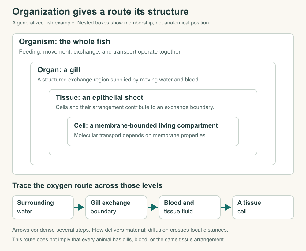
A tissue is an organized association of cells and the material around them performing related roles. An epithelium is a sheet of cells that covers or lines a surface. Its arrangement matters: a sheet can separate two spaces, direct exchanges between them, and provide a continuous boundary that isolated cells could not provide in the same way.
Muscle tissue contributes force, nervous tissue contributes communication and coordination, and connective tissues include cells within material that supports or links structures. Organs combine tissues into larger working arrangements. These categories help explain familiar animal bodies, but they should not become a requirement that every animal must reproduce one vertebrate layout. OpenStax: animal primary tissues
Imagine trying to make an exchange surface from a loose pile of otherwise suitable cells. Some might face the resource, some might be buried, and gaps might allow material to bypass the intended route. Rearranging the same cells into a supported sheet changes the problem. Orientation and connection now matter alongside each individual cell's properties. A list of cell types cannot describe that geometry.
This also explains why “made of cells” does not mean “nothing but cells.” The spaces between cells, fluid within those spaces, and material produced by cells help determine the body's operation. A route drawn straight from one cell interior to the next would miss an extracellular step if the substance actually passes through surrounding fluid. Extracellular means outside cells, which need not mean outside the animal.
Inside a body outline can still be outside a tissue
Draw a tube through the middle of a larger outline. The tube's open interior, or lumen, lies geometrically within that outline, but its contents remain separated from the surrounding tissues by a lining. In a digestive tract, a swallowed item has entered the lumen. It has not thereby entered every living cell or even crossed the tract's lining.
This distinction becomes clearer with a thought experiment. Place a colored bead in a transparent tube. You can move the bead along the tube without moving it through the tube wall. Absorption would require passage across a boundary; transport along the lumen is a different operation. The bead is only a geometric analogy, not a model of how a particular nutrient is absorbed.
Use three colors in your diagram: one for the lumen, one for the lining cells, and one for the fluid beyond the lining. Then draw an arrow that crosses from the lumen into a lining cell and another that exits toward the internal fluid. Those crossings identify questions about the two sides of the cell. A line that jumps over the lining hides the work it must explain.
The same spatial discipline helps with respiratory structures. Water or air can move through a passage near an exchange tissue without becoming the fluid bathing every cell. Ventilation moves the environmental medium. Exchange moves particular substances across the separating surface. Confusing those steps would suggest that a fish simply fills its muscle spaces with seawater whenever it breathes.
Trace oxygen to a working cell
For a generalized gill-breathing fish, begin with dissolved oxygen in water moving past a gill surface. Oxygen crosses the thin separating tissues into blood and is transported toward other tissues. At those tissues, oxygen must leave the transport route and cross the remaining distance to cells. The gill is an exchange interface within a longer supply pathway. OpenStax: systems of gas exchange
Our map deliberately omits detailed gill architecture and blood chemistry, which the fish course will develop. Nevertheless, it already distinguishes several possible limitations: bringing water to the surface, crossing that surface, moving blood, and transferring oxygen locally. Improving one step will not necessarily improve delivery if another step remains strongly limiting. “More oxygen in the environment” is not a complete prediction of tissue supply.
Think of the pathway as connected spaces and boundaries rather than a set of names. The environmental water is one space; blood is another; the fluid around a muscle cell is another; the cell interior is another. Some arrows represent movement within a space, while others represent transfer across a boundary. Marking the difference gives your diagram explanatory value even before adding detailed structures.
A useful final check is to follow the route backward from the cell. What lies immediately outside it? How does oxygen reach that location? What replenishes the transport fluid? Working backward exposes the missing final steps that an attractive drawing of a whole fish can conceal. The size of the animal does not make those last local distances disappear.
A shared cellular problem allows different body plans
The fish pathway is one arrangement, not a template into which every animal must fit. A thin aquatic body may keep exchange surfaces close to its cells. A larger body may place extensive surfaces within itself and connect them through transport routes. An insect's respiratory tubes present another organization: the route for oxygen differs from the blood-centered route just described. The later courses will identify examples and exceptions in detail.
At this stage, compare diagrams by the problem they solve. Where is the external medium? How far is the receiving cell from it? What moves over long distances, and what crosses a short boundary? Which structures maintain the route? These questions allow comparison without pretending that similar functional needs imply identical organs or a single best body plan.
Consider two imaginary animals made from the same number of identical cells. Arrange one as a thin sheet and the other as a compact block. More cells in the block are separated from the outer surface by other cells. The comparison changes access geometry even though the total cell count remains the same. Chapter 3 will put numbers on related size-and-distance problems.
The sheet is not automatically a superior animal. Its arrangement also affects protection, support, movement, and exposure. A useful biological comparison follows the consequences of a change instead of scoring complexity. What is convenient for one exchange problem can make another task harder, depending on the environment and the animal's activity.
Give every arrow a material and a destination
Return to the original claim that the fish obtains oxygen from water. You can now expand it into a route with specified spaces, boundaries, and kinds of movement. The improvement is not simply a longer sentence. It is a set of places where the explanation could be checked or where a changed condition could alter the result.
Apply the same discipline to a nutrient. Begin with the form available outside a receiving cell, and distinguish any required processing from movement along a route. If a large food particle is broken into smaller molecules, that is a chemical or mechanical processing step, not merely relocation. If a molecule crosses a membrane, state that separately from its later transport elsewhere.
A complete map also has exits and transformations. Cells do not only receive material. They use, alter, release, and store it. Drawing inward arrows alone makes a living body resemble a container that fills indefinitely. Adding an outward route or a specified transformation makes the account obey the quantities it claims to describe.
You do not need to know every transporter to notice a missing boundary. You do need to avoid filling the gap with a word that merely repeats the outcome. “Absorbed” identifies an event to explain. The next question is which material crossed which surface, in which direction, and through what available mechanism. That question will remain useful from a jellyfish's tissue to a mammal's intestine.
Application
Draw two interiors
Draw a simplified animal outline containing an open digestive tube and a separate tissue cell. Label the tube lumen, the lining, extracellular fluid, and cell interior. Trace a hypothetical absorbable nutrient from lumen to the receiving cell, marking boundary crossings differently from movement within a fluid. Treat the drawing as a functional schematic rather than a particular species' anatomy.
Repair the oxygen route
A student's diagram reads: “Water → fish → muscle.” Replace it with a sequence for the generalized gill-breathing fish described above. Include at least two exchange locations and distinguish water movement from internal transport. Then name one piece of information the diagram still lacks.
Check the balance
An invented cell starts with 40 units of a substance. During one minute, passive entry contributes ten units, outward transport removes six, and a reaction consumes three. Assume no other inputs, outputs, or changes of rate within that minute. How much remains? Explain why a steady amount in a different example would not imply an inactive cell.
Model interpretation
The tube lumen and the receiving cell interior are different spaces. The nutrient must cross the lining's route to reach internal fluid, then reach and enter the receiving cell. A more detailed account would identify the nutrient and the relevant transport mechanisms; the schematic alone does not establish them.
The oxygen map follows environmental water past a gill, exchange into blood, internal blood transport, exchange toward local tissue fluid, and entry into the receiving cell. It does not show exact exchange distances, oxygen binding, flow patterns, or species-specific ventilation. Naming one of these limits is better than inventing a universal number.
The balance is 40 + 10 − 6 − 3 = 41 units. The amount rises by one unit even though much more material moves or is consumed. A steady amount could instead occur when total input balances output and consumption. It would describe a net balance, not the absence of molecular activity.