A layered organ
An ink mark, a shallow abrasion, and a deep cut can occupy the same patch of a hand. From above, all three appear to change its surface. In depth, they involve very different relationships. Ink may remain outside the living tissue. An abrasion can remove part of the epidermal covering. A deeper cut can interrupt connective tissue, small vessels, and sensory structures as well as the surface boundary.
The purpose of an anatomical section is to reveal those relationships. Skin is not a uniform wrapping whose functions disappear in proportion to the area removed. It is an organ made of several tissue types, with structures reaching across named levels. The same surface area can therefore participate in protection, exchange of heat, sensation, and repair through different arrangements beneath it.
Begin at the outside and choose a direction
Superficial means closer to the body's surface; deep means farther from it. These directions are useful even when a person turns a hand over or lies down. They do not mean upward and downward in the room. In this course's sections, the external environment appears at the top, but that is a drawing convention rather than an anatomical rule.
The epidermis is the outer epithelial component. Beneath it, the dermis is connective tissue containing cells, extracellular matrix, blood vessels, and many associated structures. The hypodermis, also called subcutaneous tissue, lies deeper. It connects the skin with underlying structures and includes varying amounts of adipose and connective tissue.
Some introductory resources describe all three as skin layers. A stricter anatomical usage reserves skin proper for epidermis and dermis and describes the hypodermis as underlying tissue. We will use that distinction while including the hypodermis in our diagrams. Otherwise, a disagreement over naming can be mistaken for a disagreement about where a structure actually lies.
The NIAMS introductory skin lesson groups the organ's functions around protection, sensation, temperature regulation, and other contributions. Our next step is to assign those contributions to structures rather than imagine that every cell performs every task. You can revisit the shared anatomical orientation for epithelium, connective tissue, blood flow, and diffusion.
The epidermis is a changing population
The epidermis is keratinized stratified squamous epithelium. Stratified means it has multiple cell layers. Squamous describes the flattened cells toward the surface, not the shape of every cell throughout its depth. Keratinized refers to specialization involving keratin proteins and the formation of a protective cornified surface.
Most epidermal cells belong to the keratinocyte lineage. Keratinocytes are living cells in the deeper epidermis, where they produce structural proteins and participate in signaling and barrier formation. As they differentiate toward the surface, they change shape and composition. Their final cornified form, a corneocyte, has lost its nucleus and most organelles and becomes part of the outer barrier.
That transformation makes two common descriptions incomplete. Calling skin dead ignores its living epidermal cells, connective tissue, vessels, nerves, and appendages. Calling every surface cell alive ignores the specialized corneocytes that perform an important protective role after terminal differentiation. An organ can incorporate structures that no longer maintain the usual activities of a living cell.
Keratin is a family of structural proteins, not a liquid coating poured over the body. Within cells, keratin filaments help distribute mechanical stress through connections to other structural elements. The mature surface also depends on a tough cell envelope, intercellular lipids, and connections between corneocytes. Chapter 2 will explain why keratin alone cannot account for permeability.
Follow the epidermal strata without mistaking them for separate organs
The stratum basale is the deepest epidermal stratum, adjacent to the basement-membrane region. It includes proliferating keratinocytes and their progenitors. New cells help maintain the population as other cells differentiate and are eventually shed. A basal keratinocyte is still a keratinocyte; basal position does not place it outside that lineage.
The stratum spinosum contains keratinocytes connected by strong junctions called desmosomes. In prepared tissue, the connections can contribute to a spiny appearance as cell outlines retract. The appearance helps histologists recognize a region, but it should not be imagined as sharp external spikes on living skin. Preparation changes how tissue is seen.
The stratum granulosum contains differentiating cells with conspicuous keratohyalin granules. Among the processes occurring here are organization of keratin-associated material and delivery of lipids that help establish the surface barrier. Keratohyalin granules and lipid-containing lamellar bodies are distinct structures. A diagram that combines them into one generic granule hides different contributions.
The stratum corneum is the outer cornified layer. Its flattened corneocytes are embedded in organized extracellular lipid. Cells are eventually shed through regulated changes in their attachments, while replacement arrives from deeper epidermis. The overall surface is maintained through an ongoing process rather than a once-built sheet that merely wears away.
Thick palmar and plantar epidermis also has a recognizable stratum lucidum, between the granular and cornified strata. It is not a separate layer equally visible everywhere. Likewise, a fixed count of cells or a universal number of days for complete turnover would make the account falsely precise. Body site, age, condition, and measurement method influence such descriptions.
The boundary between epidermis and dermis is an interface
The epidermis rests on a basement membrane, a specialized extracellular interface that supports attachment and organization. The junction often has interlocking contours: downward epidermal ridges meet upward dermal papillae. These shapes increase contact and help resist mechanical separation, while bringing small dermal vessels near the avascular epidermis.
Avascular means lacking blood vessels. Blood does not normally circulate through capillaries inside the epidermis. Living epidermal cells receive supplies through exchange from the dermal vascular network and diffusion through the intervening tissue. A vessel close to the epidermis is not thereby located within it. This distinction matters when explaining why some superficial damage does not bleed.
For an original scale exercise, imagine a drawing in which a 0.1-millimeter layer is enlarged to 20 millimeters. The linear magnification is 200 times. A nearby 1-millimeter region would need to appear 200 millimeters thick at that same scale. If an illustrator compresses it to fit the page, the image may remain useful but is no longer uniformly scaled. Relative function cannot be inferred from the apparent thickness of colored bands.
The OpenStax account of skin layers provides a regional starting point. Here, the crucial relationships are avascular epithelium above vascular connective tissue, attachment at an interface, and variation across sites. The original course diagrams simplify dimensions while preserving those relationships.
The dermis combines support with a route for supplies
The superficial papillary dermis is relatively loose connective tissue. The deeper reticular dermis contains more densely arranged collagen bundles. These are connected regions rather than two sheets separated by a perfectly sharp wall. Their arrangement helps the skin tolerate forces arriving from different directions.
Collagen contributes tensile strength: resistance to being pulled apart. Elastic fibers support recoil after deformation. The surrounding hydrated matrix, cells, and fiber organization also affect behavior. A material's mechanical performance depends on arrangement as well as the names of its ingredients. More collagen alone does not guarantee that repaired skin will behave exactly like the original.
Fibroblasts are connective-tissue cells that produce and remodel extracellular matrix. They operate alongside immune cells, vessels, nerves, and other residents. The dermis therefore does more than hold the epidermis in place. It supplies a living environment in which mechanical support, communication, and defense can change over time.
Arterial branches, capillary networks, venous drainage, and lymphatic vessels form routes through the dermal and deeper tissues. Capillaries support exchange; larger vessels deliver and remove blood; lymphatic vessels return fluid and participate in immune traffic. A red and blue pair in a simple drawing represents these networks, not two pipes that visit every cell directly.
Appendages complicate a simple stack of layers
A hair follicle is an epithelial structure extending into deeper tissue around the developing hair. Its origin from the epidermal system does not mean its entire adult extent lies within the superficial epidermis. The follicle's living cells and associated connective tissue support production of a hair shaft, while the exposed shaft itself consists largely of keratinized material.
Sebaceous glands commonly open into hair follicles and release sebum, a mixture containing lipids and cellular material. Their secretion contributes to the local surface environment. They are not evenly distributed over every body site, and the absence of a hair in one microscopic section does not establish that the original region had no follicles at all.
Eccrine sweat glands include a coiled secretory portion deeper in the skin or subcutaneous tissue and a duct leading to the surface. That duct crosses levels on our diagram. A gland's location and its opening are different anatomical facts. The gland may secrete fluid beneath the surface while the visible result appears at a small pore above.
Apocrine sweat glands occur in selected regions and commonly connect with hair follicles. They differ from the widespread eccrine system, so the two should not be collapsed into one universal sweat-gland type. Their names are historical anatomical terms; a name alone is not sufficient evidence for the exact cellular mode of secretion.
Nails are another epidermal specialization. Living cells in the nail-producing region form a hard plate that extends beyond them. As with hair, visible hard material and the tissue producing it must be distinguished. That principle allows us to reason about appendages without treating every visible projection as a separate living organ.
A microscopic slice can cut the same structure several times
The Yale skin laboratory emphasizes that an epidermal derivative can occupy the dermis or deeper tissue. This is especially helpful when interpreting gland profiles. A thin section through a coiled tube can show several separate round or oval shapes. They may be parts of one continuous gland rather than several independent glands.
Imagine slicing a loosely coiled length of tubing with a flat plane. If the plane crosses five loops, the section contains five openings. Counting the openings as five entire tubes would overestimate the number of structures. Conversely, a plane passing beside the coil could miss it even though it lies nearby in the three-dimensional tissue.
The same issue affects hair follicles, nerves, and blood vessels. Their appearance changes with the angle and depth of the cut. A transverse profile may look circular while a longitudinal profile looks elongated. Identification therefore combines relationships, cellular appearance, and section orientation. No external specimen image is reproduced in this course; the exercise concerns how to interpret the limitations of sections in general.
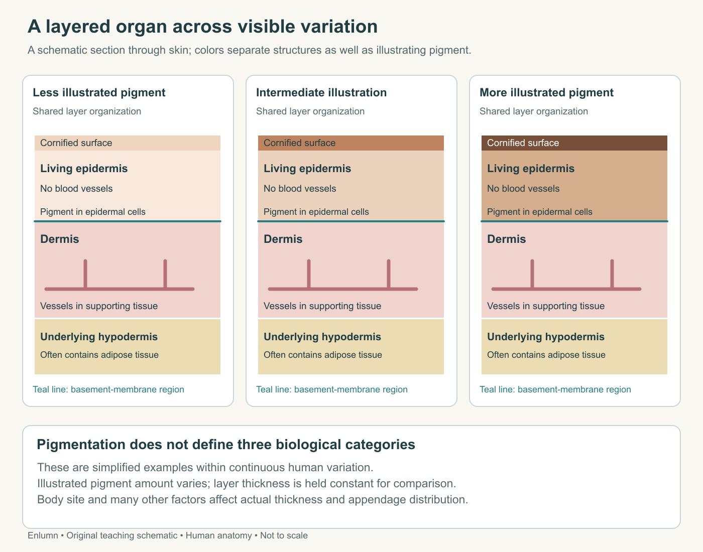
Pigmentation varies within a shared anatomical organization. The panels are teaching schematics, not racial categories or uniformly scaled specimen sections.
Regional variation is part of the anatomy
The palm and a hairy forearm are not identical skin with different decorations. Palmar skin has a conspicuously thick epidermis and lacks hair follicles and sebaceous glands. Eccrine glands remain important there. Hair-bearing regions have a different arrangement of appendages and generally thinner epidermis. Both are functioning skin.
In anatomical usage, thick skin chiefly refers to the specialized epidermis of palms and soles, not a universal ranking of the total thickness or toughness of every body region. A region can have substantial dermis without belonging to that histological category. Everyday meanings of thick, tough, and sensitive should therefore be separated from the terms used to identify tissue.
Subcutaneous tissue also varies. Adipose tissue stores energy and contributes to insulation and cushioning, but its depth and arrangement differ around the body. A standard section with a large yellow bottom band does not establish that every site has the same adipose layer. Nor does pigmentation tell us the thickness of that band.
Let depth explain the consequences
Return to the three marks on the hand. Surface ink may alter appearance without interrupting tissue continuity. An epidermal abrasion can disturb the barrier while leaving dermal blood vessels intact. A deeper injury can interrupt the vascular supply and connective framework as well as the epithelial surface. Whether a specific injury bleeds depends on what was actually damaged, not merely on its visible width.
This gives us a method for the remaining chapters. Locate the affected structure, identify its contribution, and trace consequences through nearby relationships. Barrier loss concerns the organized boundary. Altered sensation concerns receptors and neural routes. Heat exchange involves circulation and sweating. Repair must rebuild continuity and support using whatever tissue and cells remain available.
Check your understanding: A coiled sweat-gland profile appears below the epidermis, but its duct opens at the surface. Is that a contradiction? Explain why a shallow epidermal injury might affect the barrier without bleeding.
Expected answer: No. Epidermal origin and adult location are different: an appendage can extend into dermis or subcutaneous tissue while its duct reaches the surface. The epidermis lacks blood vessels, so damage confined there can disturb barrier structure without directly cutting a dermal vessel. Actual consequences depend on depth and structures involved.
Application
Spend about 15 minutes drawing a skin section from outside to subcutaneous tissue. Label the epidermis, dermis, basement-membrane region, a dermal vessel, hair follicle, eccrine gland and duct. Give superficial and deep directions. Mark an invented abrasion confined to epidermis and another injury extending into dermis.
Write one paragraph explaining two different consequences of depth. Then draw three oval profiles and explain why they might represent one coiled gland rather than three glands.
A successful answer keeps blood vessels outside the epidermis, lets appendages extend across levels, distinguishes skin proper from underlying tissue, and avoids using surface area alone to predict injury. It explicitly states that a two-dimensional section does not reveal every three-dimensional connection.