A long organ with a large working surface
A forkful of pasta has disappeared from sight, but most of its useful material has not yet entered your circulation. Swallowing moved it into a passage. The stomach mixed it and released portions downstream. A glucose unit still locked in starch remains on the other side of a living boundary, even though it is physically within your abdomen. This distinction between location and access is the starting point for understanding the small intestine.
Imagine following two parts of that meal: a glucose unit in the pasta and a long-chain fatty acid in its olive oil. Both must reach an epithelial surface. Their chemistry will require different processing, and their routes away from the intestine will diverge. Neither can be explained by drawing an arrow labeled “food” straight into the blood. This course builds the missing middle of that arrow, from the arrangement of the organ to the behavior of its cells.
A passage with a boundary
The lumen is the space within a hollow organ. In the intestine, it contains water, digestive secretions, food-derived molecules, microorganisms and material shed from the lining. The lumen connects with the external environment through the mouth and anus. Calling it functionally outside the body's internal environment is useful, provided we remember that it is a highly regulated space surrounded by living tissue. It is not exposed to the surroundings in the same way as a tabletop.
The epithelium forms its immediate cellular boundary. On one side are intestinal contents; on the other are supporting tissues, blood vessels and lymphatic vessels. Absorption requires movement across this boundary. Digestion changes molecules into forms that can be absorbed. These processes overlap in place and time but answer different questions: what chemical form is available, and can it cross?
A dissolving substance illustrates the distinction. Sugar can disappear visibly into water while every sugar molecule remains in the glass. Likewise, disappearance of recognizable food fragments does not establish absorption. A liquid intestinal sample can contain abundant unabsorbed molecules. Visual smoothness tells us little about which molecules have reached an epithelial cell, crossed it or entered a vessel.
Use the shared anatomical orientation when reading the diagrams. Right and left refer to the person whose body is represented. Proximal and distal describe positions along the route, here closer to or farther from the stomach. These terms remain meaningful even when a loop bends back toward its starting point. A distal segment can lie physically beside a proximal one without changing their order.
Three regions along one continuous tube
The duodenum begins at the stomach's pylorus. Much of it lies relatively fixed against the posterior abdominal region and forms a curve around the pancreatic head. Here acidic gastric contents meet pancreatic secretions and bile. This proximity matters: useful digestive substances can reach the lumen near the beginning of a long absorptive passage. The organs contribute through ducts, while the food itself remains inside the intestinal tube.
The jejunum follows the duodenum. The ileum follows the jejunum and ends at the junction with the cecum of the large intestine. There is no separate door between jejunum and ileum, and their differences develop gradually. A diagram that colors them differently is identifying regions, not claiming a sharp painted boundary exists inside the body. Many nutrients are absorbed over overlapping stretches.
The jejunum generally has more prominent circular folds than the distal ileum. The ileum remains an absorptive organ and has particular importance for the uptake of the intrinsic-factor–vitamin B12 complex and for recovery of bile acids. Regional specialization does not mean that each nutrient has a single tiny assigned station. It means that some mechanisms are distributed unevenly, making location relevant when function changes.
The jejunum and ileum occupy mobile loops supported by the mesentery, a fold of peritoneal tissue connecting them to the posterior abdominal wall. Within that support run vessels, lymphatics and nerves. The mesentery therefore does more than keep an inconveniently long tube from moving about. It provides routes through which the intestinal wall receives oxygen and resources, and through which absorbed material can leave.
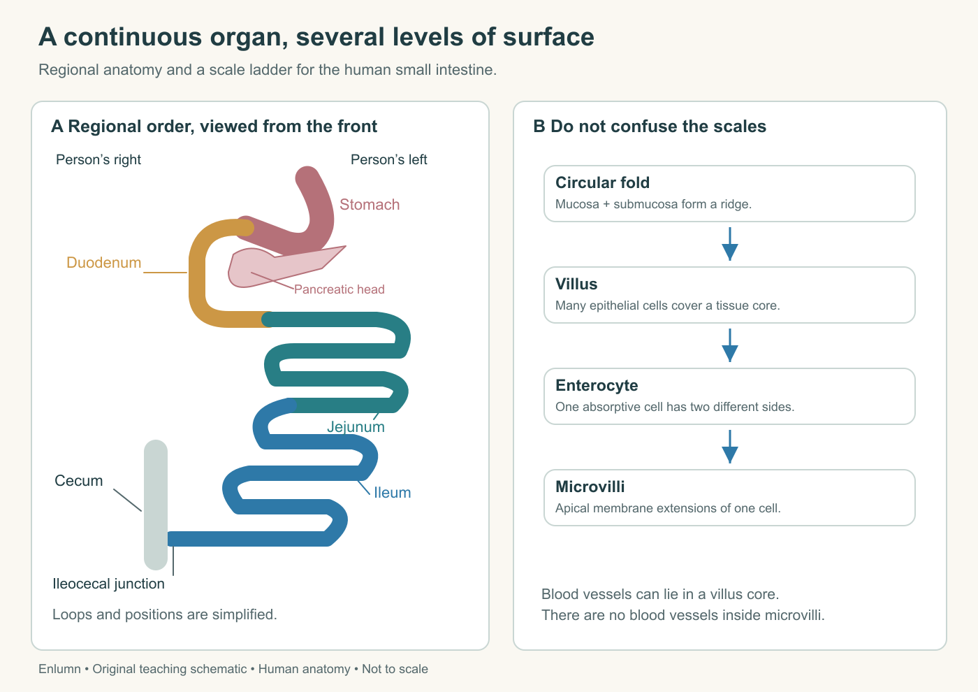
The map preserves regional order and person-relative orientation. Loop positions and proportions are schematic. The adjacent scale ladder separates whole-organ folding from villi and the microvilli of individual cells.
Surface at three different scales
A circular fold is a ridge involving mucosa and submucosa. A villus is a much smaller projection of mucosa, covered by epithelium and containing a supporting core. A microvillus is smaller again: an extension of the apical membrane of an individual epithelial cell. The prefixes and similar names can make these structures seem interchangeable. They are not.
A villus contains many cells and internal tissue. A microvillus is part of one cell and contains no blood vessel. Drawing a capillary inside a microvillus confuses scales by several orders of magnitude. Conversely, representing a whole villus as one absorptive cell loses the supporting tissue and the separate vascular and lymphatic destinations within its core.
The brush border is the collective appearance of closely packed microvilli on absorptive cells. It provides membrane area for digestive enzymes and transport machinery. The microvilli do not behave like a broom sweeping food into holes. Nor are they the motile cilia found on some other epithelia. Their relevance here is the arrangement of membrane and its molecular equipment.
Consider a sheet of paper laid flat inside a box. Folding it allows more sheet area to occupy the same floor footprint. That is a useful geometric analogy for an elaborated surface, but paper does not need a blood supply or replace its cells. Living folds must be maintained, supplied and reached by luminal material. A more elaborate surface would accomplish little if its cells lacked transporters or its crevices were inaccessible to the substances being absorbed.
Histological descriptions of the jejunum from Yale's teaching collection distinguish these levels: submucosa extends into the large folds, while the villi project from their mucosal covering. This is a useful way to read an unfamiliar section. Ask which tissues form the projection before naming it from its outline alone.
The wall supports the surface
An enterocyte is an intestinal absorptive epithelial cell. Its apical surface faces the lumen; its basal and lateral surfaces face supporting tissue and neighboring cells. The two sides contain different molecular machinery. This polarity permits coordinated transport through the cell instead of identical exchanges in every direction.
Under the epithelial layer lies the lamina propria, connective tissue containing vessels, immune cells and other supporting elements. A thin smooth-muscle layer, the muscularis mucosae, completes the mucosal organization. Beyond it lie submucosa and the larger muscle layers of the muscularis externa. These larger layers contribute to mixing and propulsion. A muscle layer outside the mucosa is not itself the membrane across which glucose is absorbed.
Crypts extend down between villi. They contain renewing cell populations and specialized secretory cells. New epithelial cells arise from stem-cell populations in the crypt region, and many subsequently move toward the villus surface as they differentiate. This renewal maintains a boundary exposed to continual mechanical, chemical and biological activity. Different cell types have different life histories; a single turnover number should not be assigned to every cell.
Goblet cells contribute mucus. Paneth cells near crypt bases contribute antimicrobial substances. Enteroendocrine cells release signals that help coordinate digestive activity. Immune structures and cells participate in responses to intestinal contents. The lining is consequently more than a uniform sheet of nutrient collectors. Its ability to absorb depends on an environment that these other cells help maintain.
How large is the working surface?
You may have heard that the intestinal surface is the size of a tennis court. That comparison is memorable, but it hides assumptions about tube length, diameter, folds and microscopic amplification. None of those quantities is constant across people, regions or measurement methods. Fixation, stretching and the condition in which length is measured also affect estimates.
Helander and Fändriks revisited this question in a 2014 human morphometric study. Their abstract describes literature review and light- and electron-microscopic measurements from human biopsies. They estimated a mean total digestive-tract mucosal area of roughly 32 square meters, including about two square meters for the large intestine. This is an estimate from an approach, not a newly discovered exact area that every person possesses. We are using the published abstract, not claiming to have inspected the full measurement dataset.
The scientific lesson is how an attractive analogy can survive after its numerical basis becomes questionable. Replacing one sporting surface with another does not teach the measurement. A useful account identifies what was measured directly, what was estimated from samples, and how local observations were scaled to an organ.
Try an original geometric example unrelated to that study's data. Suppose a smooth cylinder has an inner circumference of 8 centimeters and a length of 300 centimeters. Its lateral area is 2,400 square centimeters, or 0.24 square meters. Suppose folds multiply that by 1.5, smaller projections by 8, and cell-surface elaborations by 12. The resulting modeled area is 34.56 square meters: 0.24 × 1.5 × 8 × 12.
The multiplication is valid only if each factor describes an additional level relative to the preceding one. If a published factor already includes villi and microvilli, multiplying by a separate microvillus factor again counts that surface twice. This error can produce a precise-looking result that is wrong in its construction. Before calculating, write what each factor includes.
Area does not equal absorption
Even a correct area estimate is not a direct measurement of nutrient uptake. Imagine two fictional epithelial surfaces, each with 100 equal patches. In one, all patches transport a selected substance at two units per minute. In the other, only half have functioning transport machinery at that rate. Their anatomical areas are equal, but their modeled uptake capacities are 200 and 100 units per minute.
Now imagine the first surface receives only 60 units per minute of available substrate. Its capacity remains 200, but its actual sustained uptake cannot exceed the supplied 60 in this simplified example. Capacity describes what could occur under stated conditions. Throughput describes what does occur. Confusing the two makes every large surface seem automatically productive.
Access and removal matter too. Material must mix near the membrane. Absorbed substances must move away through tissue and vessels. Cells require metabolic support. An obstruction downstream of one of these steps can reduce overall transfer without immediately changing the visible number of villi. A structure-function explanation should therefore connect at least three elements: surface organization, cellular mechanism and supporting flow.
This also changes how we interpret a section under a microscope. A thin cut can pass through the side of a villus, making it appear as an isolated round profile. The space around it is still continuous with the lumen in three dimensions. A two-dimensional image does not show the entire villus length, the full blood network or the rate of absorption. Observing structure and inferring function are related activities, not identical measurements.
Make a map that can answer a question
Return to the pasta and olive oil. Your first map should show their shared route through the duodenum toward jejunum and ileum. Your second should zoom to a villus, with its epithelial boundary and internal support. Your third should show one enterocyte with a luminal and a tissue-facing side. Keeping these maps separate prevents a long arrow from concealing several distinct crossings.
To test the map, place a dot representing a dissolved glucose molecule in the lumen. Ask whether it is inside a cell, within tissue fluid or within blood. Move the dot only when you can name the boundary it crosses. Then place a dot within an epithelial cell and ask whether reaching that position guarantees delivery to the rest of the body. It does not; transport out and onward still remain.
Finally, ask what information the picture omits. The organ map omits microscopic structures. The cell map omits most of the body's circulation. Neither omission is a flaw if it is explicit and appropriate to the question. Trouble begins when a diagram designed to show regional order is treated as a measurement of length, or a drawing of increased area is treated as evidence of a particular uptake rate.
Check your understanding: A learner draws a blood vessel inside each microvillus and says that doubling villus height must double nutrient absorption. What needs correcting?
Expected answer: Microvilli are extensions of individual cell membranes and contain no blood vessels; vessels occupy the tissue core of villi. Greater surface dimensions do not by themselves determine uptake, which also depends on available substrate, transport machinery, access, cellular support and removal into circulation.
Application
Spend 15–20 minutes making three nested drawings: a regional intestinal route, a villus with its supporting core, and one polarized enterocyte. Mark the lumen on each drawing where it exists. Add one feature that cannot be resolved at that scale.
Then calculate the original cylinder example after changing the fold multiplier from 1.5 to 2, keeping everything else fixed. Explain why the result is not a prediction of an individual's absorption.
Model interpretation: The revised geometric result is 46.08 square meters. It changes an assumed surface multiplier, not transporter function, nutrient availability, blood flow or measurement evidence. The organ drawing should preserve duodenum–jejunum–ileum order; the villus should contain tissue; the microvillus should belong to a single cell.