A chamber with layers
A mouthful of rice and egg takes a few seconds to swallow. Its subsequent handling cannot be completed on that schedule. The meal must be mixed with secretions, its particles worked on, and its delivery coordinated with a much longer stretch of digestive tract. Between swallowing and intestinal absorption stands a chamber whose wall is both an active machine and a living boundary. Understanding that wall is our first task.
Start by setting aside the familiar use of “stomach” for the whole abdomen. The anatomical stomach is one organ. An ache somewhere below the ribs does not identify which organ produced it, and a meal has not finished digestion when it leaves the stomach. In this course we will follow structures and processes closely enough to avoid both confusions. You need no previous organ course. If anatomical directions are unfamiliar, the shared orientation in The Heart explains how body positions and sections are described.
Follow the passage, then leave it
The alimentary canal is a continuous passage from mouth to anus. Its central space is the lumen. Swallowed material travels through the esophagus into the stomach and then through the pyloric opening into the duodenum, the first part of the small intestine. Most nutrient absorption takes place farther along the digestive process, principally in the small intestine. The stomach contributes storage, processing, and delivery. NIDDK's digestive-system overview places these functions within the complete route.
Draw that route as a line. Now draw a second line through the stomach wall: from an artery into small vessels supplying the tissue and then into veins. These lines represent different journeys. A piece of swallowed rice does not normally enter the artery supplying the stomach. Oxygen carried by that artery does not arrive by being swallowed. The organ receives contents through one route and resources for its own cells through another.
This distinction changes what “inside the body” means. Food in the lumen is physically enclosed by your body, but it has not necessarily crossed an epithelial boundary into your internal tissues. An intestinally absorbed molecule has crossed such a boundary; a solid fragment sitting in the stomach has not. Imagine a tunnel through a mountain. A truck in the tunnel is within the mountain's outline without being embedded in its rock. The comparison concerns geometry only, but it prevents the assumption that enclosure itself is absorption.
There is a third relevant space outside the stomach wall: the peritoneal cavity, normally a thin potential space between serosal surfaces. It is not the gastric lumen and is not a blood vessel. A future chapter will use this distinction to explain perforation. For now, make three separate labels on your drawing: lumen, living wall, and outside surface. Every explanation of gastric protection depends on keeping them separate.
A map with a movable outline
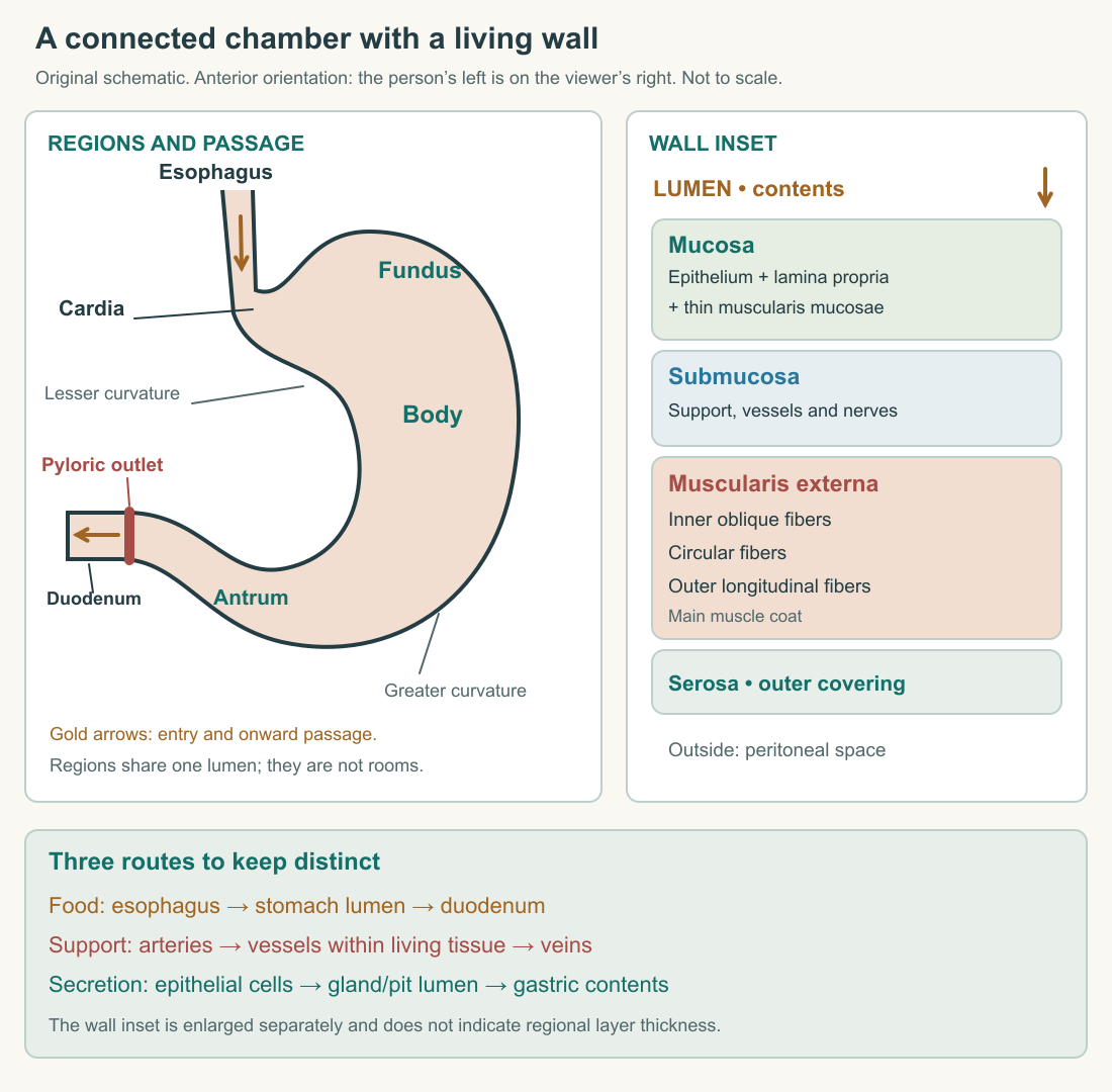
The stomach occupies the upper abdomen, largely to the left. At the esophageal entrance is the cardia. The fundus forms the dome above and to the left of that entrance, beneath the diaphragm. The larger central region is the body, also called the corpus. Toward the exit, the pyloric antrum narrows into the pyloric canal and the muscular pyloric sphincter. The longer convex border is the greater curvature; the shorter concave border is the lesser curvature. OpenStax's structural account gives this regional orientation.
These are regions of a connected chamber, not four sealed compartments. An arrow running cardia, fundus, body, pylorus can mislead if it suggests that every mouthful files through four successive rooms. The entrance joins a space whose contents can redistribute. The fundus is not a compulsory checkpoint between entry and exit. Similarly, the greater and lesser curvatures are borders, not ducts that transport different foods.
Read the original diagram in two passes. First follow the open route from esophagus to duodenum. Then look at the wall inset. The first pass is about where contents travel; the second is about what the boundary is made of. Neither drawing gives a universal organ size or a fixed position after every meal.
The liver lies partly in front of the stomach; the diaphragm arches above it; the pancreas is related to its posterior surface across the lesser sac. The spleen is a neighbor toward the left. Peritoneal folds connect the stomach with other structures: the lesser omentum reaches toward the liver, while the greater omentum descends from the greater curvature. These relations help explain how an organ can move and expand while remaining connected to vessels and neighboring tissues. The anatomical reference describes these attachments and relations.
A map should therefore preserve connections more firmly than silhouette. Consider a road map drawn on flexible cloth. Bending the cloth changes distances on the page without changing which road meets which bridge. A stomach does not behave exactly like cloth, but the mapping lesson is useful: esophagus, chamber, and duodenum remain connected as shape changes. When comparing two anatomical images, ask whether they show different filling states or viewing directions before concluding that one is wrong.
Four layers, several kinds of work
From the lumen outward, the main layers are mucosa, submucosa, muscularis externa, and serosa. The mucosa itself contains an epithelial lining, supporting connective tissue called the lamina propria, and a thin smooth-muscle component called the muscularis mucosae. The submucosa carries larger supporting vessels and nerves. The substantial outer muscle coat moves the chamber's contents. The serosa is the outer covering associated with visceral peritoneum. This layered plan is described in the stomach physiology reference.
Two names contain “muscle,” but they do not refer to the same layer. Muscularis mucosae belongs to the mucosa; muscularis externa lies outside the submucosa. On your diagram, a label pointing vaguely to “the muscle” loses information. It is like identifying both a drawer runner and a building's steel frame as metal. The material is shared; the scale and mechanical role differ.
The larger muscle coat contains fibers oriented in longitudinal, circular, and inner oblique directions. Smooth muscle can change the shape and tension of the wall without your voluntarily commanding each contraction. The arrangement supports coordinated deformation rather than the action of a blade turning inside a rigid blender. The pyloric sphincter is a thickening of circular muscle at the outlet. A narrowing produced there changes the resistance faced by contents trying to leave.
Here is an original structural test. Replace the living lining with an inert waterproof sheet while leaving the outer muscle intact. The chamber might still change shape, but it would lose the epithelial cells that secrete and maintain its luminal environment. Now restore the lining and immobilize the outer muscle. Secretion could occur while mechanical handling changed profoundly. Finally preserve both but remove their blood supply. Neither layer could continue its normal work indefinitely. These are thought experiments, not procedures. They show why “muscular bag” describes only part of the organ.
The wall also contains nerves. Local enteric networks help coordinate activity, while connections with the wider nervous system help relate it to swallowing and the body's state. A nerve in a wall is not a separate hollow pipe carrying food. On a labeled section, its location tells you where information can influence tissue, not where the meal goes. The distinction between material routes and control routes will become especially useful when we examine emptying.
Folds are not glands
When the stomach is relatively empty, its mucosa and submucosa form folds called rugae. These folds flatten as the chamber expands. At a much smaller scale, the epithelial surface dips into gastric pits, which lead into glands. A large fold and a microscopic pit are different structures. Flattening a fold does not mean that all the secretory glands disappear.
Imagine a corrugated sheet with small wells molded into its surface. Straightening the broad corrugations changes the overall shape while leaving the small wells recognizable. Again, the analogy captures scale, not living physiology. A label such as “fold” is incomplete unless we specify whether it describes the whole wall, part of the lining, or a microscopic cellular structure. This is why anatomy uses both gross images and histological sections.
In the Histology Guide human fundic-stomach slide MH 113, the named Gastric Glands view shows many elongated gland profiles packed beside one another. Some profiles are cut along their length; others appear more rounded because of the cutting angle. Darker nuclei and varying pink-purple tissue contrast interrupt the neat parallel lines of a textbook drawing. The specimen metadata identifies human tissue stained with hematoxylin and eosin. The image is linked at its owner's site rather than reproduced here.
What can that section establish? It supports the claim that the lining has organized microscopic depth and repeated glandular structures. It does not directly show the rate of acid secretion, the direction of a contraction, or how full the stomach was during a meal. A stained section is a spatial record. A movement claim needs observations over time; a chemical-output claim needs an appropriate measurement. Good anatomy becomes more useful when we state both what an image shows and what it cannot settle.
To practice reading a section, pick one pale channel and follow its visible boundary. Where the profile ends, resist drawing an imaginary continuation through unrelated tissue. The section may have left the structure's plane. If two pale spaces lie close together, proximity alone does not prove they communicate. We reconstruct three-dimensional structures from slices using repeated sections, established anatomy, and other evidence. A single attractive image is not permission to invent a connection.
The stomach wall has its own economy
The meal supplies potential resources for the organism, but the stomach's cells need resources while they work on it. Blood supplies oxygen and circulating substrates to epithelial, muscle, and other cells. Venous drainage enters the portal circulation, linking the organ's drainage with the liver. The arterial supply reaches the organ through branches associated with the celiac circulation and its curvatures. We need the two routes more than a memorized list of every vessel branch.
This arrangement creates a useful causal distinction. Increasing food in the lumen increases the workload placed on the organ; it does not automatically increase resources available inside every wall cell. A machine can be surrounded by raw material while its power supply fails. The stomach is much more adaptive than that machine, yet the distinction between workload and support remains. When we discuss tissue protection, circulation will be part of the explanation rather than decorative anatomy.
Consider three observations in an invented teaching model. First, the chamber holds more material. Second, a sample from the lumen becomes more acidic. Third, a marker appears in a vein leaving the wall. These observations answer three different questions: storage, luminal chemistry, and transfer into circulation. None can simply substitute for another. Greater volume does not by itself measure digestion, and a lower pH does not demonstrate absorption of a nutrient.
You can now improve the opening sketch. Put a boundary around the lumen, draw a gland opening into it, and place a vessel on the tissue side of the epithelium. Add muscle outside the submucosa and serosa outside that. Draw one arrow for passage toward the duodenum and another for a secretion entering the lumen. The arrows should not cross into the same destination merely because both start somewhere in the wall.
The result is an explanatory map. It gives us places to put the acid-producing cells, the protective surface, the machinery of mixing, and the routes that support them. The next chapter will fill in chemical relationships, but the basic rule is already established: specify the compartment before naming the process. That habit will prevent more mistakes than memorizing another dozen labels without connections.
One last map-reading problem helps connect the scales. Suppose an endoscopic image shows the inner surface, a gross dissection shows the outer contour, and a microscopic section shows glands. Their apparent textures differ because their viewpoints and scales differ. You would not expect the external photograph to resolve individual secretory cells, nor the microscopic field to reveal the entire greater curvature. To combine them, state which surface each image faces and where its field sits in the larger organ. This is how a set of pictures becomes anatomy: each contributes a different, explicitly located piece of the same structure.
Application
Draw the stomach in outline and label entry, fundus, body, antrum, pyloric outlet, and duodenum. Add an enlarged wall inset with its four principal layers. Use different arrow styles for swallowed material, secretion, and blood. Write 150–250 words explaining why the fundus is not a compulsory first room and why a gland is not the same thing as a ruga.
Check your understanding: A diagram shows food passing from the stomach lumen directly into a vessel in the submucosa without crossing the epithelium. What is missing, and why does it matter?
Expected answer: It omits the tissue boundary and a mechanism of absorption. Material enclosed in the lumen has not automatically entered the internal circulation; a route through the lining must be established. Blood supplying the wall and food passing through the chamber are separate routes.