Secretion and protection
An egg contains proteins. So does the wall of the stomach receiving it. If the chamber creates conditions that help digest one set of proteins, why does it not simply destroy the other? The answer involves location, controlled secretion, and continual maintenance. There is no single indestructible coating. The stomach creates a chemically aggressive working space beside tissue that actively preserves a different local environment.
Our meal will remain rice and egg, but we will follow only selected processes. A useful explanation does not have to name every molecule. It must distinguish the job of an acid from the job of an enzyme, and distinguish both from the absorption of their products. It must also place secretions on the correct side of the cells making them. We begin with the word that often hides these distinctions: digestion.
Three changes that look like one
Cutting a piece of cooked egg into smaller fragments changes its particle size. Unfolding a protein changes aspects of its three-dimensional structure. Breaking peptide bonds divides a protein chain into smaller chains. These processes can assist one another, but they are not identical. Crushing a necklace into a smaller pile does not necessarily break its string; straightening it does not separate every bead. The analogy is limited, but it distinguishes rearrangement from bond cleavage.
Acid helps denature food proteins, changing their folded structure. Pepsin is a protein-digesting enzyme that cleaves peptide bonds. Gastric chief cells secrete its precursor, pepsinogen; acid-dependent activation produces pepsin, which can contribute to further precursor activation. The stomach's protein products still require further processing in the small intestine. The pepsin physiology reference explains the precursor and the acidic conditions that support enzyme activity.
A hypothetical experiment makes the distinction concrete. Place equal amounts of a purified protein into four controlled laboratory preparations: unchanged conditions, acid alone, active enzyme under suitable conditions, and enzyme under conditions that impair its activity. Measure both folding and the lengths of the resulting protein fragments. The comparison needs those two measurements because a change in appearance does not identify which bonds have been broken. This is a paper experiment, not an invitation to handle strong acid at home.
Even a tube full of short peptides does not demonstrate absorption. Absorption requires movement across an epithelial boundary into the body. The tube has no such boundary, and the stomach is not the main site where the meal's nutrient load crosses one. Keep the sequence explicit: physical processing can improve access; chemical processing changes molecules; absorption moves suitable products across tissue. One event may promote the next without being equivalent to it.
Acid is an output of living cells
Parietal cells produce gastric acid using transport machinery in their luminal membrane. The hydrogen–potassium ATPase uses metabolic energy to move hydrogen ions toward the gland lumen in exchange for potassium; chloride also reaches the lumen through its transport pathways. Stimulation changes the availability and activity of the acid-secreting machinery, including delivery of pump-containing membrane to the secretory surface. Engevik, Kaji, and Goldenring's review describes the parietal cell as a regulated secretory cell, not a passive bag of acid.
The location of the pump matters. If you draw its output arrow into the blood instead of the gland lumen, your diagram predicts the wrong destination. If you draw acid as a substance poured into the stomach from a separate organ, you remove the gastric lining's own role. The gland is a route through which epithelial output reaches the chamber. The blood supply supports the cell making that output, but it is not the delivery duct for gastric juice.
pH describes hydrogen-ion activity on a logarithmic scale. In a simple dilute-solution comparison, pH 2 corresponds to roughly ten times the hydrogen-ion concentration of pH 3. It does not mean “twice as acidic.” Nor does one pH reading tell you how much acid the stomach has secreted over an hour. Volume, buffering, ongoing secretion, and movement of contents all affect that inference.
Consider two invented vessels. Each reads pH 3, but one contains a small volume of unbuffered solution and the other a larger, strongly buffered mixture. Adding an equal amount of base may produce different pH changes. The starting pH did not specify the quantity of base required to shift it. Food brings buffering components into the stomach, so a meal can change luminal pH while acid secretion continues. A less acidic sample immediately after food arrives need not mean that the parietal cells have stopped working.
We can express the accounting without claiming to model every reaction: the acid present in a sampled region depends on acid entering, acid consumed in reactions, acid leaving, and redistribution within the chamber. A measurement of the resulting state is not automatically a measurement of one contributing rate. This distinction will return when we examine emptying: contents remaining and contents leaving per minute are also different quantities.
Signals arrive by different routes
The secretory response is coordinated by neural, endocrine, and local signals. Acetylcholine can stimulate parietal cells. Gastrin released by G cells, concentrated in the antral region, reaches targets through circulation and promotes acid secretion, including through histamine released by neighboring enterochromaffin-like cells. Somatostatin provides inhibitory control, with acid-sensitive feedback involving antral D cells. The gastrin reference introduces the stimulatory pathway; the parietal-cell review describes the broader balance of activation and inhibition.
For the diagram, group the signals by how they reach their targets rather than memorizing an undifferentiated list. A nerve terminal releases a transmitter locally. An endocrine cell releases a hormone toward internal fluid and circulation. A local mediator acts over a shorter tissue distance. None of these control arrows should be drawn as swallowed food passing down the gland. Information and digestive material occupy different routes even when both affect the same final secretion.
A negative-feedback loop changes an output in a direction that opposes a disturbance. Suppose a model chamber becomes more acidic, activating an inhibitory signal that reduces further acid secretion. That is negative feedback. It does not promise an unchanging pH at every moment. A meal may enter faster than the response develops, and the buffering properties of its contents may change. Feedback regulates a changing system; it does not abolish change.
To test the logic, remove one arrow at a time from the model. If the inhibitory signal cannot reach its target, the chamber may continue receiving a stronger secretory drive than it otherwise would. If the pump itself cannot respond, a high stimulatory signal need not produce a high acid output. Thus measuring a hormone and measuring acid are not interchangeable. A pathway contains sensors, signals, targets, and effectors, and failure at different points produces different relationships between measurements.
Protection has depth
The surface mucus layer helps retain bicarbonate near the epithelium, supporting a less acidic local environment than that in the bulk lumen. The chemical boundary is spatial: a reading in the chamber's contents need not equal a reading at the cell surface. The mucus–bicarbonate research account describes this protective association. Mucus is not an impermeable plastic liner, and bicarbonate must be supplied rather than imagined as an inexhaustible reserve.
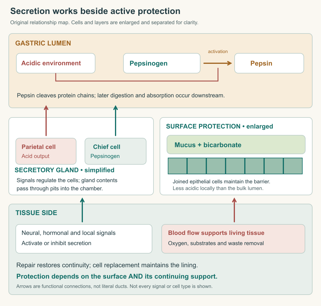
Protection also involves the epithelial barrier, rapid repair of superficial defects, replacement of cells, and blood flow that supports living tissue. Restitution refers to neighboring cells moving to restore continuity after superficial injury; it differs from making replacement cells by proliferation. The mucosal-defense review identifies these layers of defense. We are using its general mechanisms, not treating an older review as a present-day treatment manual.
The circulatory contribution deserves its own arrow. Tissue needs oxygen and substrates to maintain secretion, transport, and repair. Blood flow also helps handle substances that reach the tissue side of the barrier. Kvietys's chapter on gastrointestinal circulation and defense connects mucosal responses with perfusion. A drawing that shows only mucus leaves out the continuing support that makes the surface's work possible.
Imagine a model surface with three independently adjustable features: the acid load confronting it, the effectiveness of its protective layer, and the speed of its repair. Hold acid load constant and reduce protection. Damage can increase without acid production increasing. Restore protection but slow repair. Small injuries may persist longer. Increase acid load while keeping the other two fixed, and yet another imbalance becomes possible. This is a causal exercise, not a formula for predicting an individual's ulcer risk.
The exercise changes the question from “How strong is the acid?” to “What is the relationship between the challenge and the tissue's defenses?” It also explains why reducing a damaging influence can help even when that influence was not the original cause of the defect. Turning down water flow may limit a leak, but it does not prove excessive water pressure made the hole. We will apply this reasoning to peptic ulcers in the final chapter.
A stomach product needed elsewhere
Parietal cells also make intrinsic factor, a binding protein important for the normal uptake of vitamin B12 farther down the tract. Food-bound B12 is released with the help of gastric acid and proteolysis, binds initially to haptocorrin, and is later freed from that carrier in the duodenum. It then binds intrinsic factor; the complex is taken up in the distal ileum. The NIH Office of Dietary Supplements describes this multiorgan sequence.
This is an especially useful example because the place making an essential participant is not the place performing the final absorption. Draw four labels: release from food, temporary binding, binding to intrinsic factor, and intestinal uptake. Put them along the route rather than piling them all inside the stomach. The product of one organ can enable an event that happens somewhere else.
Now consider two hypothetical failures. In one, the relevant binding protein is missing despite food reaching the intestine. In the other, the binding protein is present but the appropriate intestinal uptake machinery is impaired. Both can disrupt the complete pathway, yet their locations differ. Merely adding the word “digestion” to both cases obscures that difference. A well-drawn route tells us which steps remain possible and which connection has been lost.
The stomach does not create the vitamin from nothing, and intrinsic factor does not turn food into red blood cells. Those shortcuts erase intake, transport, absorption, circulation, and cellular use. We include the pathway here to establish interdependence, not to prescribe supplements or interpret a blood result. Later organ courses will show the same principle in other forms: a local secretion can have consequences far beyond the tissue that releases it.
Build a meal explanation from the parts
Return to the rice and egg. Mechanical handling exposes more surfaces to the luminal environment. Acid alters protein structure and supports pepsin activation and activity. Secreted precursors and their active products participate in chemical processing. The surface maintains local protection while circulation and repair support the tissue. The resulting mixture will leave in portions for further digestion and absorption downstream.
The explanation should contain causal verbs. “Acid, pepsin, mucus, blood” is a list. “Acid supports activation of a secreted precursor; mucus and bicarbonate help maintain a protected surface; circulation supports the cells maintaining that surface” is a mechanism. To assess your own account, underline each verb and ask whether it connects the correct structures. If all your verbs are “is” and “has,” you may have named the parts without explaining their relations.
A final invented comparison separates output from success. Two model chambers secrete the same amount of enzyme. In one, mixing brings enzyme into contact with accessible protein; in the other, large intact particles limit access during the observation period. Equal secretion need not yield equal processing. Conversely, a chamber can produce a highly processed mixture and still deliver it poorly. Chemical function depends on physical handling, but a measurement of one does not fully describe the other. That is the problem the next chapter will solve.
There is also a difference between the amount of enzyme and its concentration. In a stipulated example, ten enzyme units dispersed through one hundred volume units give a different concentration from the same ten units dispersed through two hundred. That arithmetic alone cannot predict digestion, because access to substrate, reaction conditions, time, and transport also matter. It does identify a missing variable whenever someone compares two samples only by their total enzyme content. To make the comparison informative, specify what was sampled, its volume, when it was sampled, and which reaction was measured.
The same discipline applies to protection. A photograph of an apparently continuous surface is evidence about visible continuity at that moment, not a direct assay of bicarbonate secretion or future repair capacity. Functional claims require functional evidence. This does not make structural evidence unhelpful; it tells us what additional observation would strengthen the explanation.
Application
Annotate the secretion-and-protection diagram with five arrows: parietal-cell output, chief-cell output, activation in the lumen, surface protection, and circulatory support. Add a separate intrinsic-factor route ending in the ileum. Write a controlled comparison in which damage increases while acid production remains unchanged; identify what you changed and what you held constant.
Check your understanding: After a buffered meal enters, luminal pH rises even though parietal cells continue secreting acid. Is that a contradiction?
Expected answer: No. pH describes the current chemical state, whereas secretion is an input rate. Food buffering, volume, redistribution, reactions, and outflow affect the measured state, so pH alone does not establish that secretion stopped.