Locate the Parts and Supporting Structures
A person looks through a window at a tree. Light reflected from the tree reaches the eye, but the optic nerve does not carry miniature leaves to the brain. Somewhere along the route, an optical event becomes a pattern of cellular activity. Understanding vision begins by keeping those two kinds of event separate while locating the living structures that connect them.
This course follows that transformation. We will study the eye's optical arrangement, retinal signaling, differences between detail and dim-light vision, and the physiology that keeps transparent tissue usable. In the final chapter, supplied cases will show why several very different problems can produce the same everyday complaint of blurred vision. No eye disease can be diagnosed from the diagrams or exercises here.
Put the globe in its surroundings
The eyeball, also called the globe, occupies an orbit, a bony cavity containing muscles, fat, connective tissue, vessels, and nerves. The visible front is only part of the organ. Eyelids and the tear-covered surface help maintain the interface with the outside world, while the back of the globe connects to deeper structures. An eye drawn as a floating circle leaves out much of the system needed to use it.
Use the shared anatomical orientation when reading sections. Anterior means toward the front of the body, posterior toward the back. For either eye, nasal means toward the nose and temporal toward the temple. The right edge of a page is not automatically the person's right side. A front-facing illustration reverses that relationship between viewer and subject.
An adult globe is roughly two and a half centimeters across, with meaningful individual variation. That scale is small enough that structures occupying only a fraction of a millimeter can affect the optical path. If a model enlarges a 24-millimeter globe to a diameter of 240 millimeters, it is enlarged tenfold in linear dimensions. A 0.5-millimeter feature would become 5 millimeters, not 50. Cross-sectional area increases a hundredfold under the same enlargement.
Those numbers define a teaching model, not measurements of every eye. The distinction matters whenever an illustration exaggerates a thin layer to make its label readable. A diagram can preserve the order of structures while deliberately distorting their thickness. Ask what relationship it was built to show before treating it as a ruler.
Three coats, several jobs
The wall can be organized into an outer fibrous coat, a middle vascular coat, and an inner retinal region. The sclera forms much of the tough outer wall. At the front, the transparent cornea occupies a specialized part of that outer coat. Their junction is the limbus. Sclera and cornea are continuous tissues with different organization and optical roles; the cornea is not an open window cut through an otherwise complete wall.
The middle coat, or uvea, includes the choroid, ciliary body, and iris. The choroid lies between sclera and retina toward the back. The ciliary body forms a ring farther forward, and the iris extends inward in front of the lens. The iris contains pigment, vessels, connective tissue, and muscles. Its central opening is the pupil. A pupil is therefore a space bounded by tissue, not a black tissue disk.
Kolb's gross-anatomy chapter provides the three-coat and three-chamber framework. These are different classifications: coats describe the wall, whereas chambers describe internal spaces. You should be able to change from one classification to the other without adding or deleting an anatomical part.
Imagine describing a house once by its wall materials and again by its rooms. “Brick, insulation, plaster” and “hall, kitchen, bedroom” are not competing lists. Likewise, sclera, choroid, and retina do not replace the anterior chamber, posterior chamber, and vitreous cavity. Confusing the lists produces diagrams that look plausible but have impossible spatial relationships.
Trace light without sending it through every label
For a central ray entering the pupil, begin at the tear-covered corneal surface. Light passes through the cornea and aqueous fluid, through the pupil, through the lens, and across the vitreous before reaching the retina. The sequence follows an optical route. It is not a list of every tissue that supports that route.
The iris controls an opening along the route, but admitted light passes through the opening rather than through the opaque iris itself. Zonular fibers suspend the lens around its edge; they do not form a solid sheet across the central pupil. The ciliary body changes lens mechanics and produces aqueous fluid, but the central ray does not need to pass through the ciliary muscle before reaching the retina.
The National Eye Institute's explanation of how eyes work connects cornea, pupil, lens, retina, and optic nerve. We will expand the transition between the final two: photoreceptors influence retinal circuits, and retinal ganglion-cell axons form the optic nerve. Light transmission through transparent media and neural signaling along axons are different processes.
An arrow on a diagram should therefore have a stated meaning. A gold arrow might mean the direction of admitted light. A blue arrow might mean neural information moving through selected cells. Using a single unlabeled arrow for both makes it easy to imagine that photons enter the optic nerve and continue along it. The color convention is arbitrary; preserving the distinction is essential.
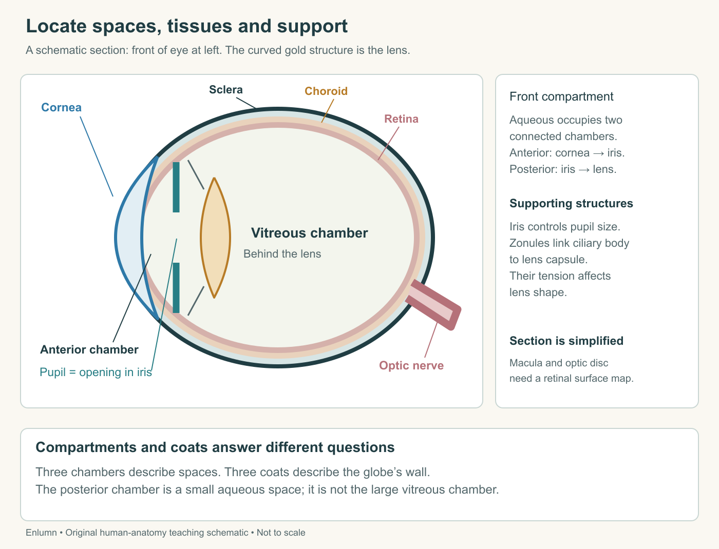
Follow the gold route before reading the labels around it. Then locate the structures that support the route without lying directly in it. This separates optical access from physiological support, two requirements we will reconnect in the maintenance chapter.
The posterior chamber is near the front
The anterior chamber lies between cornea and iris. The posterior chamber is the smaller aqueous-filled space behind the iris, bounded by the lens and associated structures. The pupil connects these spaces. Both belong to the front portion of the eye. The large cavity behind the lens is the vitreous cavity, not the posterior chamber.
The terminology can feel awkward because “posterior” sounds like it should mean the entire back of the globe. Here it names one chamber relative to the anterior chamber. Yale Medcell's eye-section description identifies the iris as the separator of the two aqueous chambers and distinguishes them from the vitreous cavity.
Test the relationships with a verbal section. Place cornea, iris, and lens from front to back. Label the space before the iris anterior chamber. Label the space behind the iris and around the front of the lens posterior chamber. Then place vitreous behind the lens. You have constructed the chamber map without needing to remember three floating definitions.
A second distinction is between chamber and segment. A source may discuss the anterior or posterior segment as a broader region containing several structures. That does not rename the posterior chamber as the posterior segment. When two diagrams seem inconsistent, check whether they use the same classification before concluding that they disagree about anatomy.
Lens support is not a set of wires carrying sight
The transparent lens sits behind the iris. Zonular fibers connect its surrounding capsule to the ciliary region. Changes in tension transmitted through this arrangement help change lens shape. The lens is not a small muscle contracting independently, and zonules are not nerves delivering an image to the retina.
Purves and colleagues' anatomical account connects the ciliary body's muscular and fluid-producing roles to surrounding structures. These roles occur in one anatomical region but involve different processes. A question about lens curvature cannot be answered merely by saying that aqueous fluid is produced, just as a question about fluid circulation is not answered by naming accommodation.
The distinction provides a useful method for learning anatomy. For each structure, write a location, an input or connection, and a contribution. For a zonular fiber, the contribution is mechanical tension transmission. For a retinal ganglion-cell axon, it is neural signaling. For a chamber, it is a space containing a particular medium. All can appear as narrow lines in a schematic, but they are different kinds of thing.
A model can also separate changes. If a lens becomes less transparent while its position remains stable, optical transmission can change without its supporting fibers breaking. If its curvature changes while transparency remains high, focus can change without clouding. These are supplied hypothetical alterations, not claims about a person's eye. They prepare us to distinguish mechanisms rather than group everything under “the lens stopped working.”
The retina has a direction and an internal organization
The retina is neural tissue with several cell populations, not a passive sheet that receives a finished picture. Rods and cones provide the photoreceptors for conventional image-forming vision. Other neurons process their signals before output leaves the eye. The retinal pigment epithelium, or RPE, lies adjacent to the photoreceptors on the outer side and performs important support functions.
Here “outer” means toward the wall and choroid; “inner” means toward the vitreous. At much of the retina, incoming light passes through inner retinal tissue before reaching photoreceptor outer segments. A signal can then progress through retinal circuitry toward ganglion cells nearer the inner surface. The optical route and the principal neural route therefore need not point in the same direction through a section.
The developmental introduction to The Retina identifies neural retina as part of the central nervous system, arising from developing brain tissue. Its location inside the eye does not make it equivalent to an inert optical material. This matters when we later compare correcting focus with repairing a neural pathway: improving the incoming image cannot automatically replace missing neural function.
Within the posterior retina, the macula is a region important for central vision, and the fovea is a specialization near its center associated with fine detail. The optic disc is a different location, where ganglion-cell axons leave the globe. It contains no rods or cones and corresponds to the physiological blind spot. The fovea and disc should never be merged into one label reading “where the eye sees.”
A section passing through the optic disc might not pass through the foveal center. Their simultaneous appearance in a simplified drawing is a teaching convenience unless the section's orientation establishes otherwise. This is another reason to treat a schematic as a map of relationships rather than a literal slice of tissue.
Moving the organ differs from focusing within it
Six extraocular muscles rotate the globe: four rectus muscles and two oblique muscles. Their coordinated activity changes where the eye points. The ciliary muscle acts within the eye's focusing apparatus. Looking toward a nearby object and focusing its image involve coordinated responses, but rotating the globe and changing lens shape remain different mechanical jobs.
A simple demonstration on paper makes the distinction. Draw an eye model pointing toward a tree and place a nearby book off to one side. Rotating the model can align its viewing direction with the book, but rotation alone does not establish that the rays from the book converge at the retinal surface. Conversely, adjusting focus while continuing to point elsewhere does not move the book into central view.
Do not learn extraocular action as “each muscle pulls the eye straight toward its name.” The geometry is three-dimensional, and a muscle's contribution depends on the eye's position. For this introductory course, coordinated rotation is the essential principle. Detailed examination of individual muscle palsies belongs beyond the supplied exercises.
The optic nerve carries visual output, while other cranial nerves carry motor commands to the eye muscles. Sharing a small anatomical space does not give them the same function. An oriented diagram that separates sensory output from motor input is more useful than one that simply labels every connecting line “nerve.”
The surface belongs in the optical system
Blinking spreads tears over the corneal surface. The tear film contributes to a smooth optical interface as well as lubrication and protection. Tears drain toward the inner corner through a drainage system leading toward the nose. They are outside the globe's wall; aqueous fluid occupies internal chambers. These fluids should not be connected as if they were one reservoir.
The NEI account of tears emphasizes that useful tears must function properly, not merely exist in a measurable quantity. We will examine that distinction later. For now, locate the tear film before the cornea and aqueous behind it. A very thin external layer can matter even though it occupies little space in a globe-sized drawing.
The anatomical map is now ready for use. When explaining vision, identify whether a claim concerns surface transmission, refraction, an aperture, a fluid space, photoreceptor activity, retinal processing, or neural output. Each term should locate a process rather than decorate a memorized list.
Check your understanding: A diagram labels the large space behind the lens “posterior chamber” and sends its light arrow into the optic nerve. What two corrections are needed?
Expected answer: The large space behind the lens is the vitreous cavity; the posterior chamber is the aqueous-filled space behind the iris near the lens. Light reaches the retina, where photoreceptors and retinal circuits generate neural output carried by ganglion-cell axons in the optic nerve. Photons do not continue through that nerve as an image.
Application
Allow fifteen to twenty minutes. Make two linked drawings from the supplied anatomy, without examining or manipulating an eye.
First draw an oriented globe section with cornea, sclera, iris, pupil, lens, ciliary body, zonules, three chambers, retina, choroid and optic nerve. Label anterior and posterior. Add separate colors for the light route and neural output, and identify at least two supporting structures not directly crossed by the central light ray.
Second draw the same globe inside a simplified orbit. Show an extraocular muscle, the eyelid and tear-covered surface. Explain in two sentences why pointing, focusing and maintaining a transparent surface are different tasks.
A strong drawing places both aqueous chambers in front of the vitreous cavity, distinguishes pupil from iris tissue, keeps tears outside the cornea and locates the optic disc separately from the fovea. At tenfold enlargement, a 0.5-millimeter model feature becomes 5 millimeters. The drawing's labels should explain relations even if its proportions are simplified.