A map of the final passage
The residue arriving from the small intestine is not finished stool. It is a fluid mixture containing unabsorbed food-derived material, secretions, microorganisms and material shed from the body itself. Much of the meal's readily absorbed nutrition has already crossed the intestinal surface, but considerable work remains. Water and ions must be handled, microbial products must be managed, material must move and storage must be coordinated with elimination.
Calling the large intestine a waste pipe captures its connection to an exit and misses most of its physiology. A pipe can conduct material without changing it or sensing its arrival. The colon does both, through living tissue supported by circulation and nerves. This course follows the changing mixture through that tissue and asks how several different processes can produce similar final appearances.
Begin at the ileocecal junction
The ileum, the final part of the small intestine, opens into the cecum, a pouch at the beginning of the large intestine. The junction helps regulate passage and limit backward movement. It is not a sorting machine that individually tests molecules and admits only useless ones. Material that passes it can still undergo chemical transformation or absorption downstream.
The appendix is a narrow extension from the cecum. It is not the route through which intestinal contents must pass to reach the ascending colon. On a route map, draw it as a side branch rather than the next compulsory station. Its wall contains substantial lymphoid tissue. That observation supports an immune association, but it should not be inflated into the claim that one proposed function completely explains the organ.
From the cecum, the ascending colon travels upward on the person's right. Near the liver, it bends at the right colic, or hepatic, flexure. The transverse colon crosses toward the person's left and bends again near the spleen at the left colic, or splenic, flexure. The descending colon continues downward on the left, followed by the sigmoid colon, which curves toward the rectum in the pelvis.
The rectum leads to the anal canal and the external opening, the anus. These are connected regions with different structural and control features. “Colon” is often used loosely for the large intestine, but an anatomical account should distinguish the colon from the cecum, rectum and anal canal when the distinction affects the explanation.
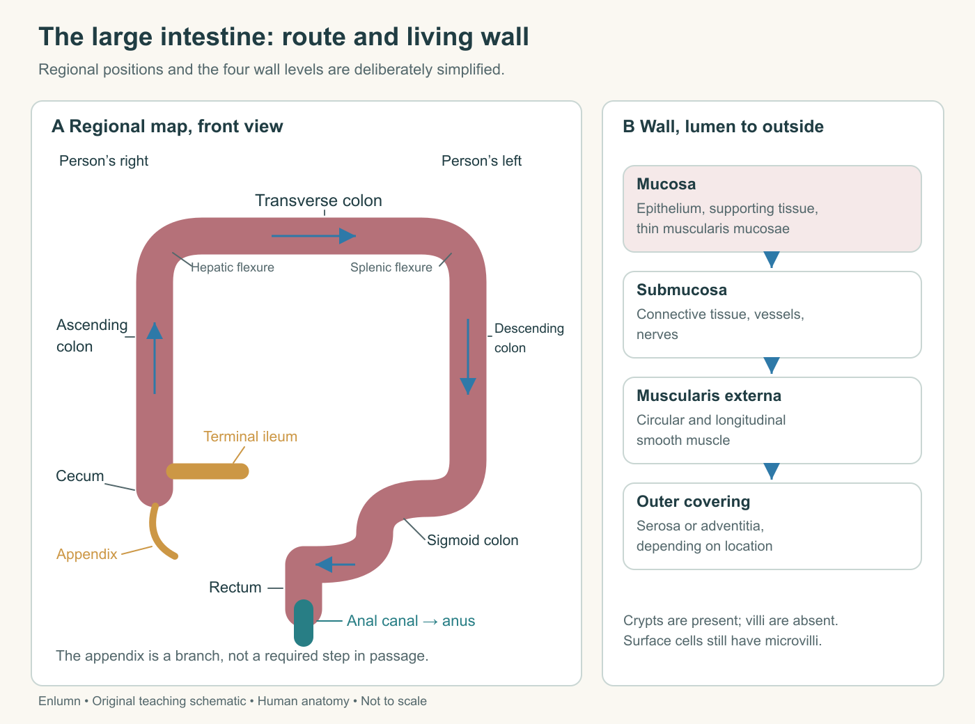
Right and left belong to the person represented. The appendix is a side branch. The diagram preserves regional sequence but simplifies the variable positions, lengths and curves of the living organ.
Names describe position, not motive
Ascending and descending describe portions of the route in the usual anatomical position. They do not mean that gravity is the main force moving contents. Coordinated muscle activity can move material through curves and along regions oriented in different directions. The transverse colon may sag, and the sigmoid colon varies substantially in shape and length. A tidy rectangular drawing is a navigational aid, not a mold into which every person's anatomy fits.
Use the shared anatomical orientation to separate position from direction of flow. Proximal means earlier along this route and distal means farther toward the anus. A proximal part can lie physically close to a distal part because the organ bends. Surgical descriptions, imaging findings and explanations of transit depend on preserving sequence despite that spatial proximity.
Much of the ascending and descending colon lies relatively fixed against the posterior abdominal wall. The transverse and sigmoid colon are supported by mesenteric folds that permit greater mobility. Those supports carry vessels and nerves; mobility does not mean a region is unattached or unsupported. The rectum's relationships change along its length and differ from those of the abdominal colon.
This regional map provides a first set of questions for a disturbance. Is the changed process early enough to affect material throughout the remaining colon? Is it close to the outlet, where a short region can influence emptying of a much larger upstream volume? Location matters through connections, not just through the percentage of organ length involved.
The wall has several jobs
The mucosa forms the inner lining. Its epithelium contacts the luminal environment and performs transport and secretory functions. Supporting connective tissue, the lamina propria, contains vessels and immune cells. A thin muscularis mucosae contributes to the mucosal organization. Beyond the mucosa lie submucosa and larger smooth-muscle layers involved in movement.
The colon's mucosal surface normally lacks the villi characteristic of the small intestine. It does have tubular crypts and absorptive epithelial cells with microvilli. Absence of villi is not absence of membrane area or absence of absorption. A different surface organization supports a different balance of tasks.
Goblet cells are abundant and contribute mucus. Other epithelial cells participate in absorption, secretion and signaling. Stem-cell populations in crypts replenish the lining. Underneath and around these cells, immune components help manage a tissue boundary exposed to a dense microbial environment. The Yale gastrointestinal histology account provides a useful description of the colon's crypt-rich, goblet-cell-rich organization.
A crypt is an inward extension of the lining, with a lumen connected to the main intestinal lumen. It is not a separate sealed gland sitting outside the digestive passage. In a thin section, a crypt cut across can look like a circular ring of cells surrounding a small opening. A longitudinal cut can look like an elongated tube. The different outlines can be different views of a similar three-dimensional structure.
An outer shape reflects muscle organization
Along much of the colon, the outer longitudinal smooth muscle is concentrated into three bands called taeniae coli. The circular muscle layer lies deeper. The colon's characteristic sacculations are called haustra. These visible pouches reflect the organization and activity of the wall rather than a sequence of independent chambers separated by doors.
At the rectum, the longitudinal bands spread into a more continuous arrangement. The rectum therefore should not be drawn as another identical haustrated colon segment. The anal canal adds specialized closure mechanisms and an epithelial transition toward a surface suited to greater mechanical abrasion. Its structure is related to controlled passage through an external opening.
Small fat-containing appendages on the colon's outer surface are called epiploic appendages. They are distinct from the vermiform appendix attached to the cecum. Similar words can mislead a learner into making one structure serve two locations. An organ map should include a label only when its position and role are clear enough to avoid that confusion.
The wall's muscle layers do not directly pull water molecules out of stool. Their movements influence distribution, contact and transit. The epithelial cells and their transport processes create conditions for water and ion movement. These functions interact, but naming them separately prevents muscle activity from becoming an all-purpose explanation.
The contents are a changing mixture
Feces include water, microbial biomass, unabsorbed material, shed cells and products of digestive and microbial processes. The mixture's composition varies with diet, transit, secretion, absorption and other conditions. It cannot be reconstructed simply by subtracting an assumed absorbed percentage from the weight of the last meal.
For an original accounting example, suppose 120 dry-material units enter a modeled region from upstream during an interval. The wall contributes 10 units, and 20 units are absorbed in forms we are counting as dry material. If there is no change in stored amount, 110 units must leave by the remaining counted routes. Whether those units are still the original food molecules is a separate question; microbial transformations can change their form.
Now include water. If 900 water units enter, 100 are secreted and 800 are absorbed, downstream water output is 200. That produces a different consistency from an otherwise identical mixture carrying 400 water units. The dry-material calculation alone cannot predict stool texture. The water balance alone cannot specify microbial composition or the original dietary source of each particle.
These figures are invented to establish the bookkeeping. Real measurements require clear definitions and sometimes additional output categories, including gases. If an experiment measures only solid output while material is converted to a gas that leaves separately, an apparent missing mass is a measurement-boundary problem, not a violation of conservation.
Food residue is not the only input
The body continually contributes digestive secretions and shed material to the lumen. Some is recovered upstream; some reaches the colon. The colon also secretes material locally. Microorganisms use available substrates, grow and release products. A stool sample therefore contains evidence of interactions among food, host tissue and microbial communities.
Consider a labeled plant carbohydrate entering the colon. Some of its carbon may remain in a polymer fragment. Some may enter microbial cells. Some may become small molecules that the host absorbs. Some may leave in gas. The label can help trace origin, but the original molecule no longer has to exist for its atoms to remain detectable.
This explains why “the body did not digest it” needs qualification. Human small-intestinal enzymes may not have hydrolyzed the compound, yet microbes can transform it later. The host may then absorb some products. That sequence differs from direct uptake of starch-derived glucose in the small intestine, even though both can contribute material the body uses.
The next chapter develops microbial fermentation without treating every microbial change as beneficial or harmful. For now, keep the categories visible: host secretion, host absorption, microbial transformation, storage and onward movement. They provide a more useful map than a single arrow from food to waste.
Circulation supports a demanding boundary
The colonic wall requires oxygen and nutrients from its blood supply. Absorption depends on viable cells, and immune defense and renewal also consume resources. Blood vessels carry away absorbed substances while supplying the tissue performing the work. The circulation supports the boundary as well as receiving its products.
Most colonic venous drainage belongs to the portal system and reaches the liver. Distal rectal and anal drainage includes connections to systemic venous pathways, so the entire final passage should not be represented as one exceptionless portal route. We do not need a vessel catalogue here; the practical lesson is that neighboring regions can have different connections.
A local problem can therefore have consequences beyond its length. Reduced perfusion can impair the cells needed for transport and barrier function. Conversely, a structurally intact surface does not establish that its circulation is adequate. A photograph of the mucosa, a measurement of blood flow and a measurement of absorption answer different questions.
This is also why the word “clean” is a poor physiological target for the colon. A functioning organ contains microorganisms and material in transit. Its task is regulated processing and elimination while protecting tissue, not permanent emptiness. Removing visible contents would not by itself establish healthy transport, motility or defense.
A map becomes useful when it predicts something
Test your regional drawing with a fictional narrowing near the outlet. Even if most of the colon remains structurally normal, movement through that short final region can constrain emptying upstream. Compare that with a localized reduction in water absorption early in the colon. Later regions may compensate to some degree, depending on their capacity and conditions. Equal lengths of altered tissue need not cause equal consequences.
Now test a histological drawing. If you remove villi from your imagined normal colon, you have changed nothing, because villi were not part of its usual surface to begin with. If you remove epithelial transport machinery while keeping the crypt outlines, the diagram can look nearly unchanged while function changes. An anatomically accurate picture prevents the first error; mechanistic understanding prevents the second.
The final passage is therefore neither a passive pipe nor a miniature repetition of the small intestine. Its regional structure, crypt-based surface, muscular organization and controlled outlet support a connected set of tasks. We will next follow water, salts and microbial substrates through that system, keeping host transport separate from microbial chemistry.
Check your understanding: Why can a short disturbance near the outlet affect elimination from a much larger part of the colon, while an equal-length change elsewhere may have different consequences?
Expected answer: Function depends on position and connections. An outlet limitation can constrain emptying of upstream contents; another regional change may affect transport or mixing and may be partly compensated elsewhere. Length alone does not specify the altered process or its effects.
Application
Spend 15 minutes drawing the route from ileum to anus. Add the appendix as a side branch, mark person-relative right and left, and distinguish colon, rectum and anal canal. Beside it, sketch one crypt opening onto a surface without villi.
Use the fictional water balance to calculate downstream output if absorption falls from 800 to 650 units while input and secretion remain fixed. Name two features of stool that this calculation does not establish.
Model interpretation: Water output rises to 350 units: 900 + 100 − 650. The result does not reveal microbial species, chemical composition of dry material, a clinical diagnosis or the exact physical consistency of a real stool.