A position between gut and body
Imagine following three things after a dinner of bread, beans and olive oil: a glucose molecule absorbed into intestinal blood, a package carrying products of fat digestion, and a bile salt that helped those products reach the intestinal surface. All may eventually encounter the liver, but they do not take an identical first route. The bread itself never travels through a liver chamber. The organ receives materials after other tissues have worked on them, changes some of those materials, and sends different outputs toward different destinations. Its position makes those relationships possible.
Find the organ before tracing its work
The liver lies mainly in the right upper abdomen, immediately below the diaphragm. It extends across the midline toward the left. Much of it is protected by the lower rib cage. In a front-facing anatomical illustration, the person's right is on your left; forgetting that convention can reverse an otherwise careful diagram. Use the shared anatomical orientation if the terms superior, inferior, anterior and posterior need refreshing.
Its broad upper surface fits against the diaphragm. Its lower surface relates to neighboring abdominal structures, including the stomach and the region of the right kidney. The gallbladder occupies a depression on the underside. These are relationships in three dimensions, not separate objects evenly spaced on a classroom poster. A drawing that pulls them apart makes connections visible while sacrificing the appearance of their actual packing.
A larger right lobe and smaller left lobe are recognizable external divisions. Additional named regions and internal vascular territories matter in detailed anatomy and surgery, but a surface boundary should not be mistaken for a wall that separates two independent organs. Blood vessels and bile ducts branch within the tissue. The IQWiG liver overview is a useful first location reference; our purpose is to use that location to explain connections rather than memorize every visible groove.
Picture a horizontal section through the upper abdomen. The liver will appear as an area of tissue whose outline changes with the height of the slice. A single round profile inside it might be a vessel cut across, not a spherical cavity. A long dark profile may be the same kind of structure cut obliquely. Always ask which plane you are seeing before identifying a structure from shape alone. This habit becomes essential when the next chapter moves from organs to cells.
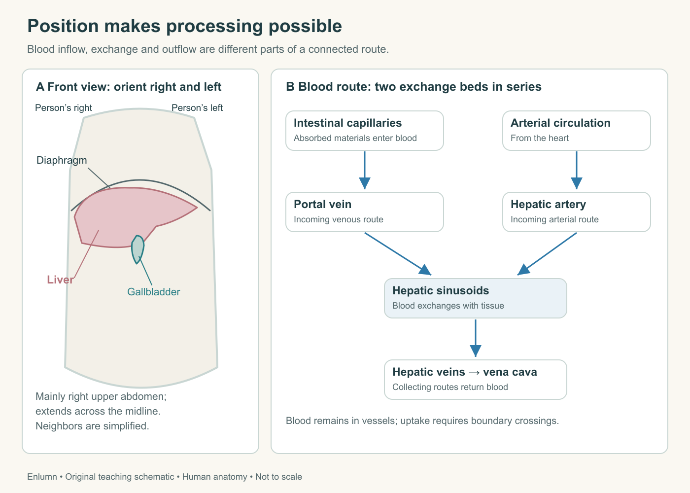
The front view uses the person’s right and left. The route diagram separates portal inflow, arterial inflow, sinusoidal exchange and venous outflow. It omits many tributaries; neither arrow width nor box size represents a measured flow or anatomical size.
Two sources of incoming blood
The liver has a dual blood supply. The hepatic artery brings blood through the arterial circulation. The hepatic portal vein brings blood that has already passed through capillary beds in digestive and related organs. The names distinguish routes, not good blood from bad blood. Portal venous blood still carries oxygen, although its oxygen content per unit volume is generally lower than that of incoming arterial blood.
The portal route is unusual because it places two capillary networks in sequence before blood returns to the heart: one in the organs being drained and another within the liver. A simplified glucose route is intestinal capillary → intestinal veins → portal vein → liver microcirculation → hepatic veins → inferior vena cava. Absorption across the intestinal lining happens before that trip through the liver. Hepatic processing happens after arrival. If the two steps are merged into “the liver digests food,” an entire epithelial barrier disappears from the explanation.
Portal blood also comes from structures such as the spleen and pancreas. It is therefore more than a delivery stream for a recent meal. It carries substances released by those tissues and materials absorbed from the gut in changing concentrations. Between meals the route remains open. Blood does not stop visiting the liver simply because the intestine has less newly absorbed glucose to contribute.
The arterial contribution supplies a smaller share of blood volume under typical resting conditions, but volume and oxygen delivery are different quantities. Oxygen delivery depends on flow multiplied by oxygen content. A smaller stream can make an important contribution if each unit carries more oxygen. Lautt's account of hepatic circulation describes the dual inflow and its convergence in the microcirculation. Exact fractions vary with physiological circumstances; the course does not turn a customary percentage into a universal constant.
Consider an original numerical model with no clinical units. Portal flow is 70 volume units per minute carrying 12 oxygen units per volume unit; arterial flow is 30 carrying 20. Their incoming oxygen deliveries are 840 and 600 units per minute. Total flow is 100, and total delivery is 1,440. The arterial route supplies 30% of volume but about 42% of the incoming oxygen. The model shows why one percentage cannot answer both questions. It is not a normal value chart for a human patient.
A shared exchange bed, a separate exit
Inside the liver, small branches of the two incoming systems supply sinusoids, specialized vascular channels running among the working cells. The cells can exchange materials with the plasma through this microvascular arrangement. They are not loose objects floating through the blood, and the entire blood stream is not forced through their interiors. Transport across a cell membrane is a selective event within a continuing flow.
Blood leaving those channels collects into small central veins and then progressively larger venous routes. The hepatic veins drain into the inferior vena cava. They are the outgoing side of the hepatic blood circulation. The portal vein is the incoming side, despite both names containing “vein.” A student who has learned that veins always leave an organ will draw this system incorrectly; vein names are defined within the circulatory route, which here includes two exchange beds in succession.
This makes the outflow as consequential as the inflow. If an outgoing route becomes difficult to traverse, the upstream system is affected. A liver with ample arterial supply would not automatically function normally if its venous drainage were severely impaired. Movement requires a pressure difference along a connected path, not merely the presence of a vessel labeled “supply.”
For a deliberately simple hydraulic model, let flow equal pressure difference divided by resistance. With a difference of 6 invented pressure units and resistance of 2, flow is 3 volume units per time unit. If resistance doubles while that pressure difference stays fixed, flow falls to 1.5. Maintaining the original flow would require a difference of 12. Real liver circulation can adjust through several mechanisms and includes parallel channels; the equation is a way to identify what must change, not a complete prediction of portal pressure.
It also clarifies why downstream and upstream observations belong together. A higher pressure before a resistant region does not mean blood moves more freely through that region. Pressure may rise precisely because passage is more difficult. Later, when we discuss scar tissue, this relationship will explain how a change within the liver can affect vessels and fluid distribution outside it.
Bile travels in another system
Bile is a fluid secretion formed by hepatocytes, the liver's principal working cells, and modified as it travels through bile ducts. It supports intestinal handling of fats and provides an exit route for selected substances. It is not the venous blood remaining after the liver has extracted nutrients. Blood and bile occupy distinct connected spaces with different boundaries and destinations.
At the smallest scale, bile enters tiny channels between neighboring hepatocytes called bile canaliculi. These connect onward to ductules and larger ducts. The right and left hepatic ducts contribute to the common hepatic duct. The cystic duct connects the gallbladder to this route; beyond that junction, the common bile duct carries bile toward the duodenum. The OpenStax accessory-organ section provides a gross duct map to compare with your own drawing.
The gallbladder stores and concentrates bile; it does not manufacture the liver's original secretion. Bile can pass from the liver toward the intestine without first entering the gallbladder. When bile is stored, flow through the cystic connection goes toward the gallbladder. When the gallbladder contracts and releases its contents, the direction through that connection reverses. A single permanently outward arrow on the cystic duct would conceal half of its role.
At the microscopic scale, blood travels from incoming portal regions toward central venous drainage, while bile travels from hepatocytes toward the duct system associated with portal regions. The phrase “opposite directions” is useful at this scale. It does not mean two straight pipes run alongside one another from the whole organ's top to bottom. Both networks branch through three-dimensional tissue. Their separation is more fundamental than whether arrows point left or right on a particular page.
What does “first pass” actually mean?
The portal arrangement exposes the liver to many absorbed substances before they enter the wider systemic circulation. This is the anatomical basis of a first-pass effect: a proportion of a substance can be taken up or transformed during its initial passage through the intestine and liver. The proportion depends on the substance and conditions. The liver does not completely remove everything that arrives, and first passage does not mean last passage.
The distinction becomes clear with an invented tracer. Suppose 100 labeled units have crossed the gut and entered portal blood. During the initial liver passage, 30 are retained or transformed and 70 leave in hepatic venous blood. It would be wrong to report that only 70 were absorbed: absorption happened earlier. It would also be wrong to claim that the remaining 30 left the body. They might be stored, converted into another molecule, returned to blood later or secreted into bile. The endpoint must be specified.
Now imagine the same 100 units arrive in systemic blood through another route. They may encounter other tissues before entering the liver through its circulation. Their eventual hepatic handling can still be substantial, but the order of exposure differs. This is an anatomical comparison, not a medication instruction. Real dose-response questions require the properties of a specific compound and its routes of absorption and elimination.
Our dinner's fat package illustrates an additional boundary. Many products of long-chain dietary fat digestion are packaged by intestinal cells into chylomicrons, which initially enter intestinal lymph. Lymph rejoins the venous circulation before these particles take their subsequent routes through tissues and the liver. Not every absorbed nutrient therefore takes the portal vein as its first route. The NIDDK digestive overview distinguishes intestinal uptake, blood transport and lymphatic transport; the small-intestine course develops that comparison in detail.
Track a material without losing its identity
A useful liver diagram must identify both the compartment and the substance being followed. “It goes into the liver” is often too vague. Does “it” mean a blood cell that remains inside a sinusoid, a dissolved glucose molecule entering a hepatocyte, or a bile salt crossing from the hepatocyte into a canaliculus? Each statement could be true while describing a different boundary crossing.
Try the bile salt from the opening dinner. It leaves the liver in bile, enters the intestinal lumen, and can later be reabsorbed into portal blood and taken up by the liver again. The returning substance does not travel backward through the common bile duct. It returns by a vascular route after crossing the intestinal surface. This circulation through intestine and liver is called enterohepatic circulation. A cycle can reuse a molecule while blood and bile remain separate fluids.
A cycle also need not be perfectly closed. Some material leaves in feces, and newly synthesized bile acids replenish the pool. If an original model sends out 100 units, recovers 95 and loses 5, the liver must add 5 to restore the starting pool for the next identical round. The total handled over several rounds can greatly exceed the size of the pool. We will return to this difference between stock and throughput when discussing bile production.
Finally, trace the organ's dependencies outward. The intestine supplies absorbed materials, the heart maintains circulation, the lungs support oxygenation, the pancreas contributes hormonal information, and the kidneys eliminate some products released into blood. Calling the liver a processing organ is useful only if processing remains connected to those other tasks. It neither receives whole meals nor independently finishes every route it begins.
Check your understanding: Why can a blood-vessel obstruction and a bile-duct obstruction have different effects even when both occur near the liver?
Expected answer: The vessels supply or drain the hepatic circulation, whereas bile ducts carry a separate secretion toward the intestine. A vascular obstruction can alter perfusion and upstream pressures; a duct obstruction can impair bile delivery and excretion. They may interact over time, but the initial compartment and disrupted route differ.
Application
Allow 12–15 minutes. Draw a route map with intestinal capillaries, portal vein, hepatic artery, sinusoids, hepatic veins and inferior vena cava. On the same page, use a distinct line style for canaliculi, hepatic ducts, cystic duct, gallbladder, common bile duct and duodenum. Show both directions through the cystic connection.
Trace three labeled journeys: absorbed glucose entering portal blood; a blood cell remaining within the vascular system; and a bile salt leaving in bile and returning through the intestine and portal circulation. Add a lymphatic route for an intestinal chylomicron. Your drawing succeeds if no arrow silently converts blood into bile or sends the contents of a meal through a liver chamber.