From nose to alveolus
Air entering your nose has not yet reached the surface that transfers oxygen into blood. It must pass through a changing route: larger passages that condition and distribute it, smaller branches whose walls differ from the trunk, and finally regions built around close contact between air and circulating blood. The lung is the place where those routes meet, but the air and blood remain in separate spaces.
This course follows that distinction from anatomy to function. Moving air is ventilation. Transferring gases across a barrier is exchange. Carrying oxygen onward depends on blood and circulation. We begin with the map because a misplaced route produces a mistaken explanation before any equation is written.
Put the lungs in a chest, not on an empty page
The lungs occupy the two sides of the chest, with the mediastinum between them. That central region contains the heart and other structures. Below lies the diaphragm, the muscular partition separating thoracic and abdominal cavities. The right lung normally has upper, middle and lower lobes; the left has upper and lower lobes and a cardiac notch accommodating part of the heart's position. NHLBI: respiratory anatomy.
Use the person's right and left, not the viewer's. In a front-facing drawing, the person's right appears on your left. A label that changes sides when you rotate the page is following the page rather than the anatomy. The shared human-anatomy orientation introduces this convention along with tissue, blood flow, diffusion and feedback; it is a reference for this series rather than a lesson repeated in every course.
The lung's apex is its uppermost region, while its base rests against the diaphragm. Its costal surface faces the ribs; its medial surface faces the central chest. Fissures separate lobes, and lobes contain smaller bronchopulmonary segments supplied by their own segmental airways. A lobe is a region of lung tissue, not one giant hollow chamber. OpenStax: gross lung anatomy.
An original mapping exercise helps establish scale. Draw the chest outline, then place the two lungs and heart. Inside one lobe, draw several branching routes. Zooming into the lobe should reveal more structure, not an empty compartment. If your drawing ends with a single balloon inside each lobe, the next stages of the map still need to be added.
The upper route conditions incoming air
Air can enter through the nose or mouth. The nasal route presents moist, vascular surfaces and projecting conchae that increase contact with the lining. Warming, humidification and particle handling begin before air reaches the chest. The pharynx provides a passage behind the nasal and oral cavities, leading toward the larynx and lower airway. Parts of this region also participate in swallowing, so neighboring routes must be distinguished. OpenStax: conducting passages.
Do not draw a direct opening from the mouth into a lung. Follow the shared throat region and then the airway through the larynx. The esophagus is a different tube leading toward the stomach. Their proximity explains why a simple line labeled throat can be insufficient when a question concerns where material travels.
The phrase conducting zone describes passages whose main respiratory role is moving and conditioning air rather than providing the principal gas-exchange surface. Conducting does not mean biologically inactive. The wall can contain living secretory, sensory and defensive machinery even where oxygen is not being transferred into pulmonary blood at an alveolar surface.
Imagine an original building plan with an entrance corridor leading to a workroom. Air reaching the entrance is necessary but not sufficient for the work occurring farther inside. The corridor can also alter what arrives there. The analogy concerns sequence and processing; a respiratory lining is living tissue, not a passive hallway.
A supported trunk becomes a branching tree
The trachea descends from the larynx and divides into right and left main bronchi. Cartilage supports the larger airways. Within the lungs, bronchi divide into lobar and segmental branches, then progressively smaller routes. Bronchioles lack the cartilage plates characteristic of bronchi and have walls whose smooth muscle can alter their caliber. NCI SEER: bronchial tree.
The names are easiest to learn as relationships. Main bronchi connect the trachea with the two lungs; lobar bronchi supply lobes; segmental bronchi supply smaller territories. Terminal bronchioles mark the end of the conducting sequence, not the end of every structure in the lung. Respiratory bronchioles begin the transition into regions with alveoli in their walls.
The word tree can mislead if we imagine perfectly repeated branches. A useful schematic omits many generations and variations. It preserves which structures connect, while exact lengths, angles and diameters require more detailed evidence. The two main bronchi also differ in their orientation and dimensions; a symmetrical drawing should not be read as proof that the two sides are identical. Amador, Weber and Varacallo: bronchial anatomy.
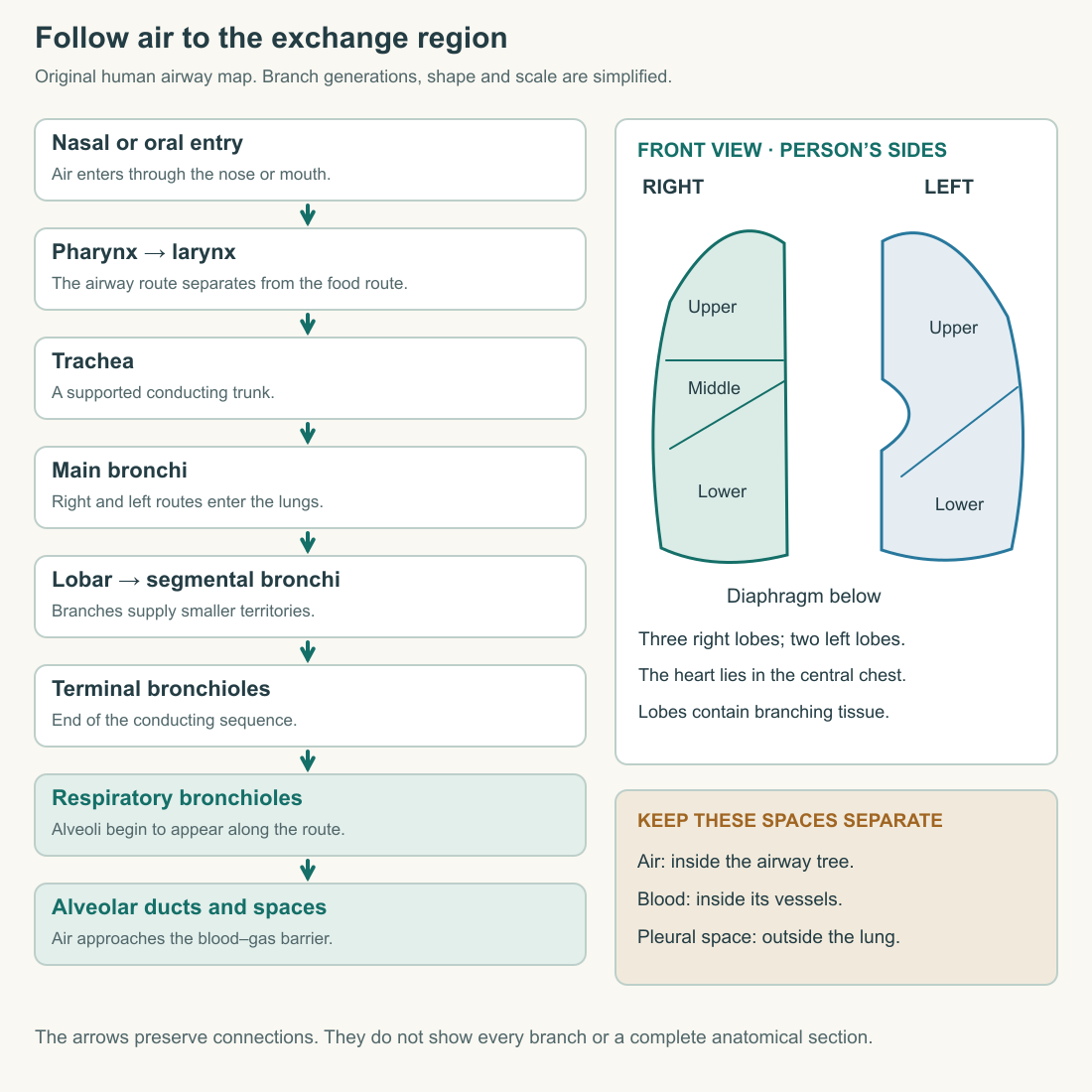
Follow one complete path on the map before surveying the whole tree. You should be able to point to a place where blocking a branch would affect all of its downstream branches. Then choose a different route that does not pass through that branch. The exercise turns a list of names into a network with consequences. Keep your finger on the continuous route as you explain those consequences.
Smaller individual branches can create more total area
A single airway becomes narrower as you follow many of the branches outward, but the number of parallel paths increases. Individual diameter and their combined cross-sectional area are therefore different quantities. Looking only at one small branch can conceal the geometry of the entire branching level.
Use an original numerical tree. A parent passage has a cross-sectional area of eight units. It divides into four passages, each with area three. Each child is smaller than the parent, yet the combined area is twelve. If the same total volume passes through the parent and the children per second, its mean speed across the combined child area is lower in this idealized account.
This calculation assumes steady flow, no leaks and equal distribution among identical children. A real breath is time-dependent, the branches are not all identical, and flow division depends on their properties. Still, the arithmetic exposes the error in assuming that narrower individual branches must mean a smaller combined route.
The distinction will matter when we discuss resistance and ventilation. A statement about one airway cannot automatically be applied to every airway operating in parallel. Structure at one scale and structure at the network scale answer related but different questions.
A human tissue slide shows the changing walls
The Histology Guide's MH 139 specimen is identified as human lung stained with hematoxylin and eosin. Its named Bronchial Cartilage view shows a substantial purple-stained cartilage region beside a folded airway lining. The Terminal Bronchiole #1 view instead shows a smaller, irregular lumen with a cellular wall amid many neighboring air spaces. The Alveoli #1 view shows stained septa separating large clear spaces. These selected views were inspected for this course. Histology Guide: human lung slide.
The slide is useful precisely because it looks less tidy than a diagram. A branching tube cut through at an angle does not necessarily produce a round outline. A folded lining can make the open space appear star-shaped. The section shows a slice through a three-dimensional arrangement, not the full length of each passage.
You do not need to identify every cell to notice the change in architecture. Compare the proportion occupied by a substantial airway wall with the much finer partitions around the smaller air spaces. Then return to the schematic and ask which details it deliberately leaves out.
Stain colors are part of the preparation. They are not a photograph of the colors of a living, ventilating lung. Nor can a fixed section show the direction of airflow or the timing of a breath. The image supplies structural evidence; movement and exchange require additional reasoning and measurements.
The exchange region has walls worth examining
Beyond the terminal conducting route, respiratory bronchioles lead into alveolar ducts and connected alveolar spaces. Alveoli are closely associated with capillaries, bringing an air-facing surface near flowing blood. Thin type I epithelial cells provide much of that surface; type II cells secrete surfactant. The barrier also includes capillary endothelium and supporting structures. It is not merely an opening between air and blood. OpenStax: respiratory zone and alveolar cells.
The distinction between a wall and an opening is fundamental. Oxygen can cross a thin barrier by molecular diffusion while red blood cells remain inside the vessel. Drawing the capillary as a pipe emptying blood into an alveolus would replace gas exchange with an entirely different event.
An alveolar wall is also shared tissue within a network, rather than the thick rubber shell of an isolated balloon. The surrounding structure supports capillaries and connects adjacent regions mechanically. A grape-like sketch can help locate small spaces, but it should not become a literal model of independent grapes hanging freely from stalks.
Chapter three will examine the barrier in detail. For now, label three places without merging them: the air space, the intervening tissue and the capillary blood. This simple separation is enough to prevent several later misconceptions about what ventilation can accomplish.
A large number is not a universal constant
Descriptions of lungs often quote a striking alveolar count. Such numbers summarize measurements made with particular methods and samples; they should not be treated as identical anatomy in every person. Ochs and colleagues used a stereological approach to estimate alveolar number in six adult human lungs. Their abstract reports substantial variation and a relationship with lung volume. Ochs and colleagues, 2004: abstract.
The methodological point is more useful here than memorizing one total. A flat section contains profiles of three-dimensional structures. Counting the visible outlines in one picture is not automatically equivalent to counting every alveolus in the whole organ. A justified sampling and reconstruction method must connect the observations with the estimate.
For an original analogy, imagine estimating the number of rooms in a building from several slices through its floors. A large room might appear in more slices than a small one. Without a rule for handling such differences, apparent abundance could reflect size and sampling as well as the actual count.
The lung study's abstract does not teach all of stereology, and this course does not claim to reproduce its full method. It establishes why quantitative anatomy needs a method and why variation belongs in the answer. The functional explanation should remain sound even when a population's average differs from a familiar textbook number.
The pleura is outside the air route
Visceral pleura covers the lung surface; parietal pleura lines the inner chest wall and related boundaries. Between them lies a normally very thin fluid-containing space. These surfaces can slide during breathing while maintaining their close mechanical relationship. The pleural space is distinct from the air-filled passages inside the lung. OpenStax: pleural arrangement.
Draw two nested outlines around a simplified lung and label the interval between them. Now trace inhaled air again. Its normal path stays inside the airway tree; it does not first enter the pleural space and then seep through the lung's outer surface.
This distinction prepares the pressure chapter. Pressure within alveoli and pressure around the lung are different measurements. Confusing their locations can reverse an explanation of how the chest and lung move together. The labels should be in place before the pressure arrows are added.
Add the blood route without drawing it into the airway
Blood reaches the pulmonary capillaries from the right side of the heart through the pulmonary arterial route and returns toward the left side through pulmonary veins. Oxygen exchange occurs along the capillary interface; air is not pumped through the heart. NHLBI: breathing and blood flow.
The lung's own living tissues also need a supply. Bronchial arteries belong to the systemic circulation and contribute blood to conducting tissues and other supporting regions. This is distinct from the much larger pulmonary route serving gas exchange. Amador, Weber and Varacallo: bronchial circulation.
That distinction changes how we interpret the word function. The lung helps supply other tissues while being tissue that must itself be maintained. A diagram can emphasize its exchange circulation for clarity, but the omitted supporting supply has not ceased to exist. Marking the omission is more useful than adding every small vessel to a crowded introductory picture.
A complete map now has two routes approaching an interface and one separate surrounding compartment. You can follow air, follow blood, and locate the pleural space without jumping from one into another. That is the anatomical foundation for explaining a breath: not a memorized collection of labels, but connected spaces and living boundaries with different jobs.
Application
Draw the airway route from nasal entry to an alveolus, then add a separate blood route and the two pleural layers. Label the person's right and left, identify the lobes, and mark where the conducting zone gives way to the respiratory region. Your drawing may simplify shape but must preserve connections.
Open the linked MH 139 human slide. Compare its named Bronchial Cartilage, Terminal Bronchiole #1 and Alveoli #1 views. Write 250–350 words separating visible features from inferred function. If the viewer is unavailable, use the descriptions in this chapter and state that you worked from supplied descriptions rather than inspecting the image yourself.
Finally, calculate the combined area of six invented branches, each two area units, compared with their parent of nine units. Explain why a smaller individual diameter does not determine total area or prove an exact real-lung flow pattern.
Check your understanding: Does air entering the trachea mean it has already reached the gas-exchange surface, and does it normally pass through the pleural cavity?
Expected answer: No. The trachea belongs to the conducting route, which leads through smaller branches to respiratory regions. Inhaled air stays within that route; the pleural space lies outside the lung between its coverings and the chest-side lining. Air, blood and pleural space must remain distinct on the map.